

• 311448
| Product name | B-hDLL3 MC38 |
|---|---|
| Catalog number | 311448 |
| Strain background | C57BL/6 |
| Aliases | delta like canonical Notch ligand 3, SCDO1 |
| Tissue | Colon |
| Disease | Colon carcinoma |
| Species | Mouse |
| Application | B-hDLL3 MC38 cells have the capability to establish tumors in vivo and can be used for efficacy studies. |
on this page
The mouse Dll3 gene was replaced by human DLL3 coding sequence in B-hDLL3 MC38 cells. Human DLL3 is highly expressed on the surface of B-hDLL3 MC38 cells.
Gene targeting strategy for B-hDLL3 MC38 cells. The exogenous promoter and human DLL3 coding sequence were inserted to replace part of murine exon 2 and all of exon 5. The insertion disrupts the endogenous murine Dll3 gene, resulting in a non-functional transcript.
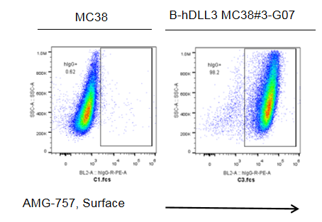
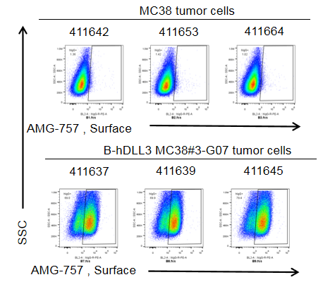
DLL3 expression analysis in B-hDLL3 MC38 cells by flow cytometry. Single cell suspensions from wild-type MC38 and B-hDLL3 MC38 cultures were stained with anti-human DLL3 antibody AMG-757 (in house). Human DLL3 was detected on the surface of B-hDLL3 MC38 cells, but not on the surface of wild-type MC38 cells. The 3-G07 clone of B-hDLL3 MC38 cells was used for in vivo experiments.
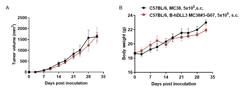
Subcutaneous tumor growth of B-hDLL3 MC38 cells. B-hDLL3 MC38 cells (5x10⁵) and wild-type MC38 cells (5x10⁵) were subcutaneously implanted into C57BL/6 mice (female, 7-week-old). Tumor volume and body weight were measured twice a week. (A) Average tumor volume ± SEM. (B) Body weight (Mean± SEM). Volume was expressed in mm3 using the formula: V=0.5 X long diameter X short diameter2. As shown in panel A, B-hDLL3 MC38 cells were able to establish tumors in vivo and can be used for efficacy studies.
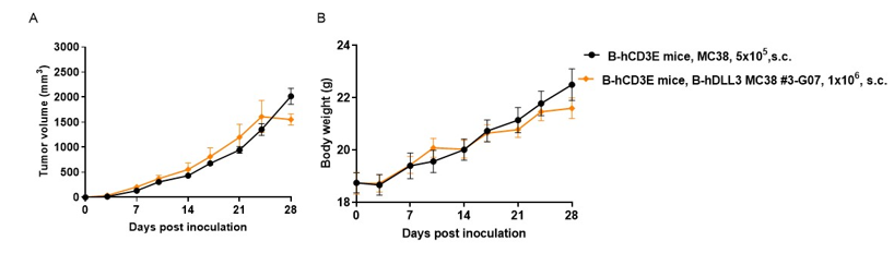
Subcutaneous tumor growth of B-hDLL3 MC38 cells. B-hDLL3 MC38 cells (1x10⁶) and wild-type MC38 cells (5x10⁵) were subcutaneously implanted into B-hCD3E mice (female, 7-week-old, n=6). Tumor volume and body weight were measured twice a week. (A) Average tumor volume ± SEM. (B) Body weight (Mean± SEM). Volume was expressed in mm3 using the formula: V=0.5 X long diameter X short diameter2. As shown in panel A, B-hDLL3 MC38 cells were able to establish tumors in vivo and can be used for efficacy studies.
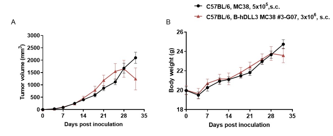
Subcutaneous tumor growth of B-hDLL3 MC38 cells. B-hDLL3 MC38 cells (3x10⁶) and wild-type MC38 cells (5x10⁵) were subcutaneously implanted into C57BL/6 mice (female, 8-week-old, n=8). Tumor volume and body weight were measured twice a week. (A) Average tumor volume ± SEM. (B) Body weight (Mean± SEM). Volume was expressed in mm3 using the formula: V=0.5 X long diameter X short diameter2. As shown in panel A, B-hDLL3 MC38 cells were able to establish tumors in vivo and can be used for efficacy studies.
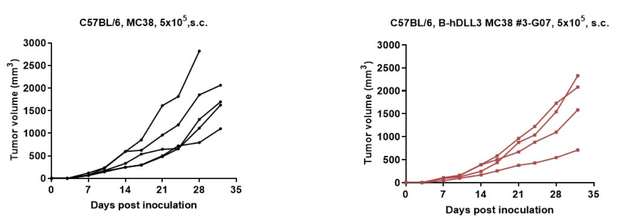
B-hDLL3 MC38 tumor cells growth of individual mice. B-hDLL3 MC38 cells (5x10⁵) and wild-type MC38 cells (5x10⁵) were subcutaneously implanted into C57BL/6 mice (female, 7-week-old). As shown in panel, B-hDLL3 MC38 cells were able to establish tumors in vivo and can be used for efficacy studies.
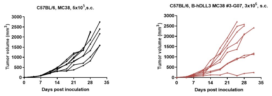
B-hDLL3 MC38 tumor cells growth of individual mice. B-hDLL3 MC38 cells (3x10⁶) and wild-type MC38 cells (5x10⁵) were subcutaneously implanted into C57BL/6 mice (female, 8-week-old, n=8). As shown in panel, B-hDLL3 MC38 cells were able to establish tumors in vivo and can be used for efficacy studies.
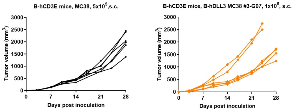
B-hDLL3 MC38 tumor cells growth of individual mice. B-hDLL3 MC38 cells (1x10⁶) and wild-type MC38 cells (5x10⁵) were subcutaneously implanted into B-hCD3E mice (female, 7-week-old, n=6). As shown in panel, B-hDLL3 MC38 cells were able to establish tumors in vivo and can be used for efficacy studies.
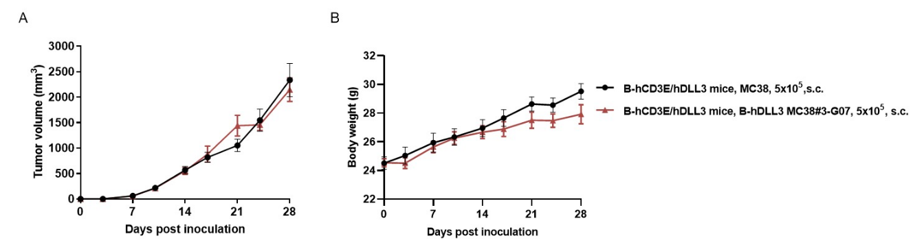
Subcutaneous tumor growth of B-hDLL3 MC38 cells. B-hDLL3 MC38 cells (5x10⁵) and wild-type MC38 cells (5x10⁵) were subcutaneously implanted into heterozygous B-hCD3E/hDLL3 mice (male, 7-week-old, n=6). Tumor volume and body weight were measured twice a week. (A) Average tumor volume ± SEM. (B) Body weight (Mean± SEM). Volume was expressed in mm3 using the formula: V=0.5 X long diameter X short diameter2. As shown in panel A, B-hDLL3 MC38 cells were able to establish tumors in vivo and can be used for efficacy studies.
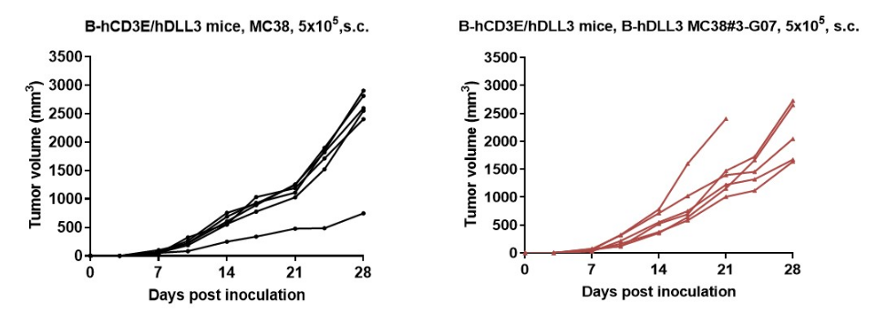
B-hDLL3 MC38 tumor cells growth of individual mice. B-hDLL3 MC38 cells (5x10⁵) and wild-type MC38 cells (5x10⁵) were subcutaneously implanted into heterozygous B-hCD3E/hDLL3 mice (male, 7-week-old, n=6). As shown in panel, B-hDLL3 MC38 cells were able to establish tumors in vivo and can be used for efficacy studies.
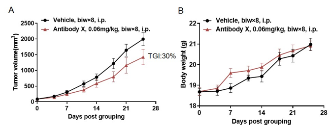
Antitumor activity of bispecific antibody (BsAb) X in B-hCD3E mice. B-hDLL3 MC38 cells were subcutaneously implanted into B-hCD3E mice (female, 8 week-old, n=6). Mice were grouped when the tumor size was approximately 100 mm3, at which time they were treated with BsAb X provided by the client with doses and schedules indicated in panel. (A) Tumor volume changes during treatment. (B) Body weight changes during treatment. As shown in panel A, BsAb X was efficacious in controlling tumor growth in B-hCD3E mice, demonstrating that the B-hCD3E mouse model is a powerful tool for in vivo efficacy study of T cell bispecific antibody. Values are expressed as mean ± SEM.
