
Biocytogen's pathology platform offers advanced capabilities in pathology diagnosis and analysis, toxicology studies, biomarker analysis, and histopathological analysis of different species and disease models, all designed to support your drug discovery efforts.
on this page
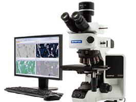
Safety concerns are one of the main 3 reasons for drug failure during clinical trials. To address this, early-stage investigative non-GLP toxicology studies are crucial, providing valuable translational information that allows pharmaceutical companies to better assess risks and benefits during drug development. Biocytogen’s pathology platform offers advanced capabilities in pathology diagnosis and analysis, toxicology studies, biomarker analysis, and histopathological analysis of different species and disease models, all designed to support your drug discovery efforts. Our platform features fully automated equipment for blood routine and biochemical analysis, high-throughput tissue sectioning and tissue microarray preparation, comprehensive staining techniques including H&E, IHC, IF, and multicolor immunofluorescence, as well as the HALO AI data analysis system. These capabilities facilitate efficacy evaluation, toxicological mechanism of action studies, and clinical translational research, providing comprehensive insights throughout the drug development process.
Our pathology platform provides a comprehensive range of services, including:

9 pathologists, 10 senior technicians and 2 project manager
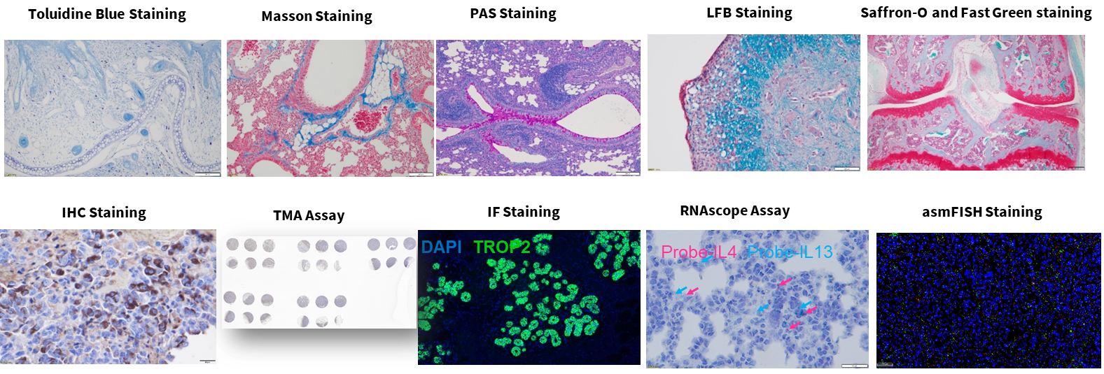
One of the most commonly used methods for paraffin and frozen sections. It facilitates pathological diagnosis while using auxiliary methods to ensure diagnostic accuracy and integrity. The nucleus, cytoplasm, and cartilage are stained blue-black, pink, and blue, respectively.
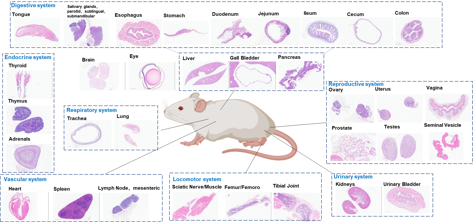
Representative H&E staining of mouse tissues
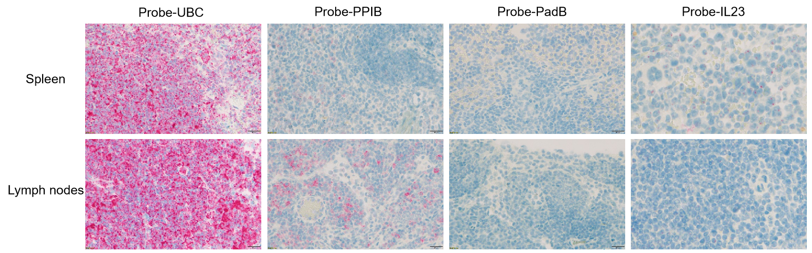
Single-stain detection analysis of RNAscope assay in psoriasis tissues
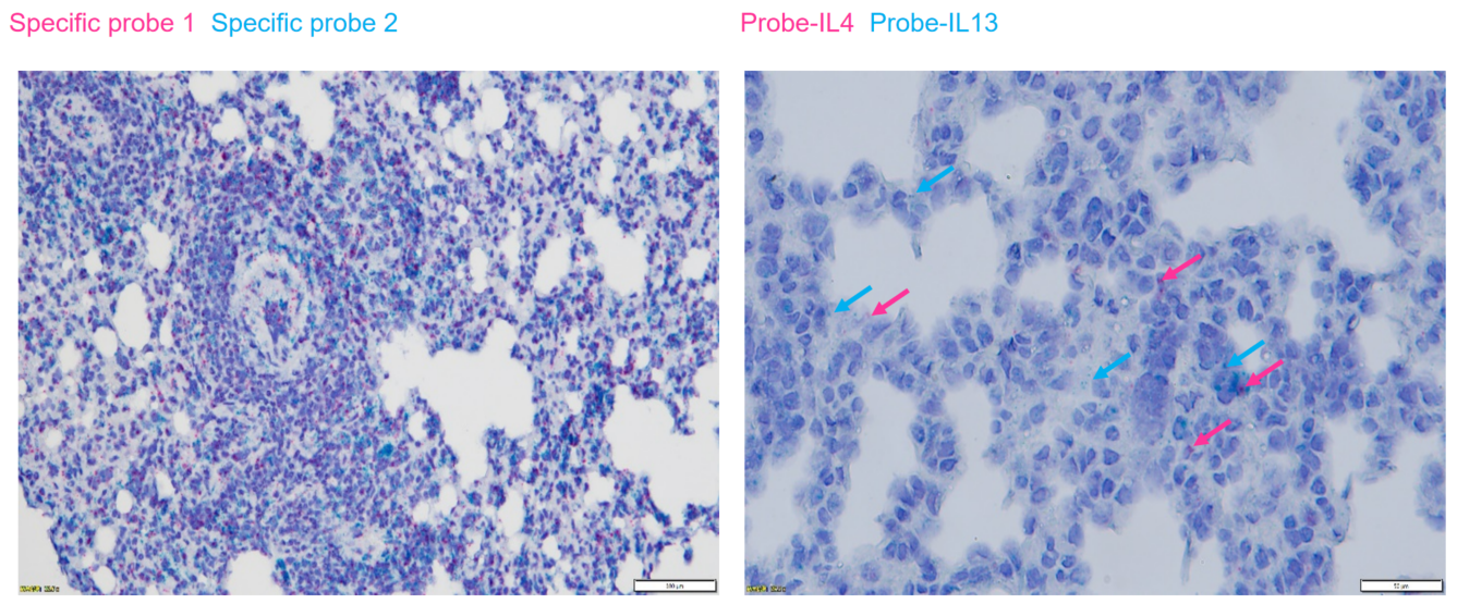
Double-stain detection analysis of RNAscope assay in asthma lung tissues
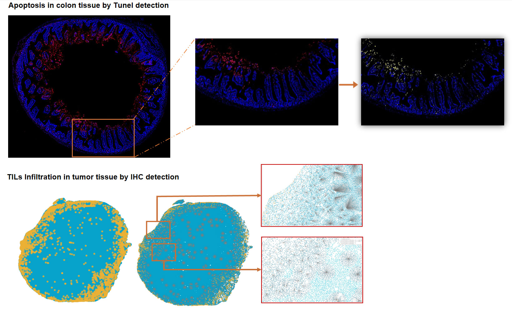
IHC/Tunel Staining and Analysis-HALO Analysis
IHC Staining and Analysis-HALO Analysis
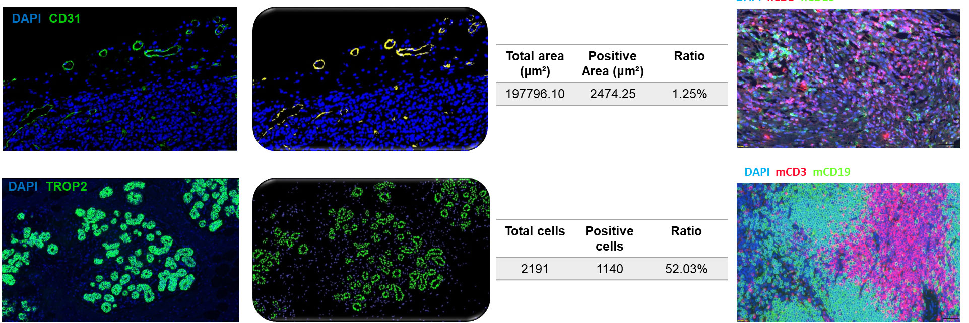
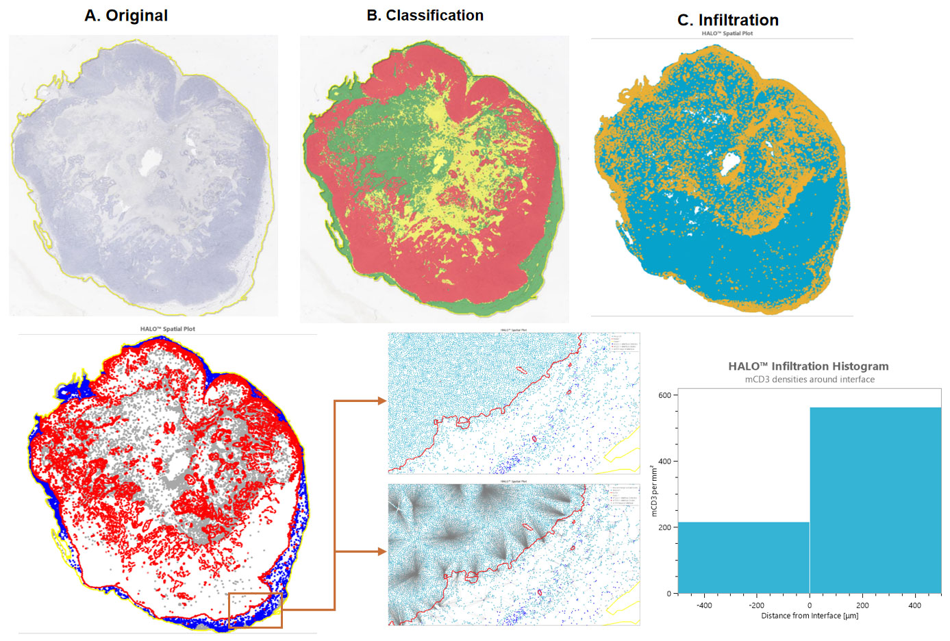
IF/TSA assays-HALO Analysis
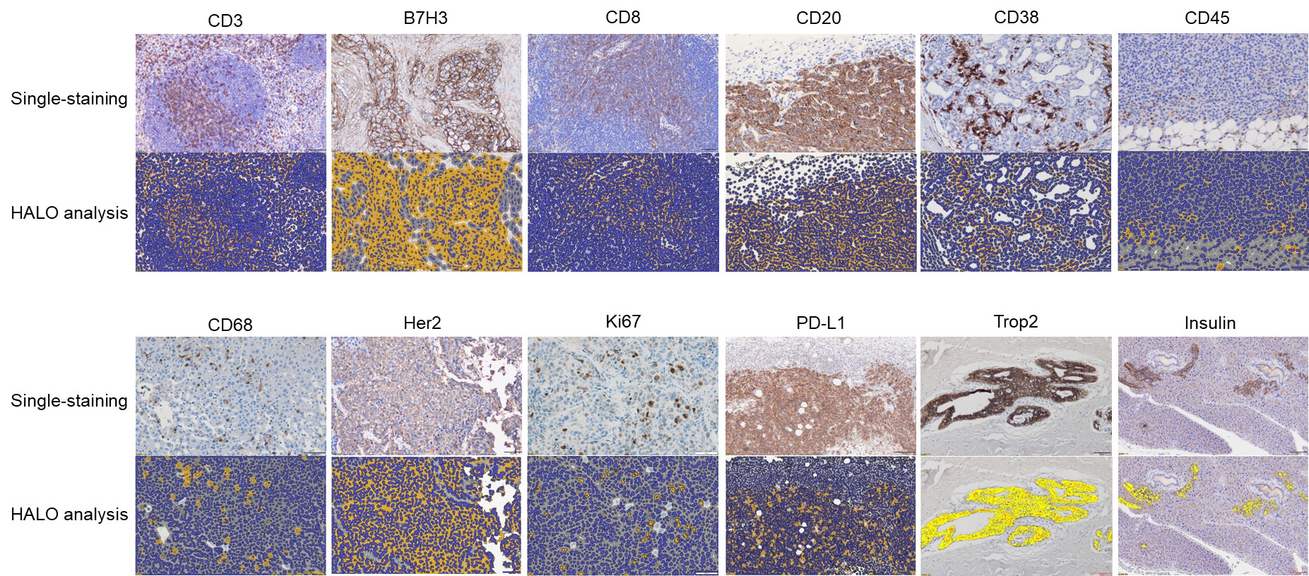
IHC assays-HALO Analysis
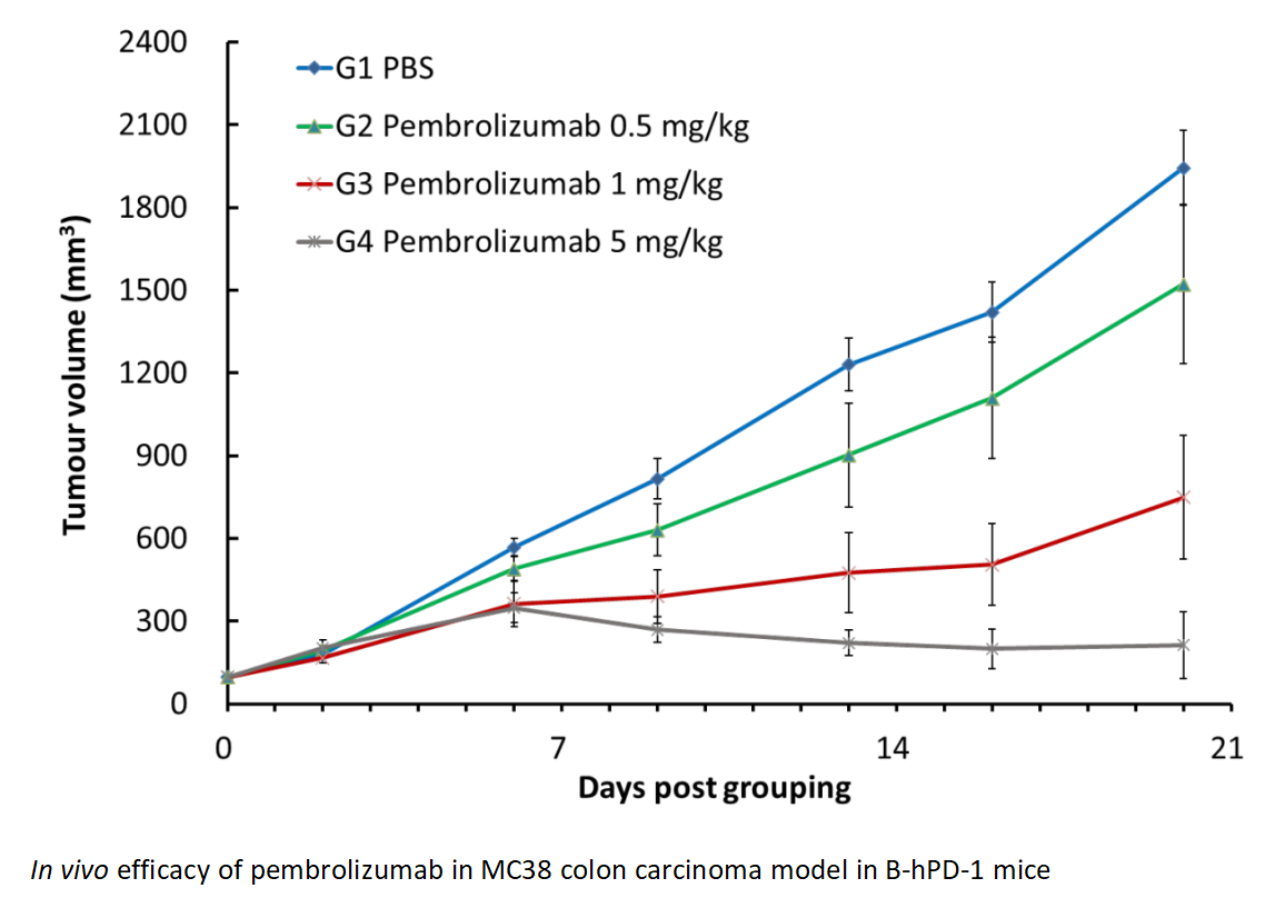
Pembrolizumab demonstrated a significant anti-tumour effect in a dose-dependent manner in MC38 syngenic model in B-hPD-1 mice. Reduced PD-1 expression in macrophages was seen in G4 compared to control, indicating macrophages are critical effector cells in anti-PD-1 treatment.
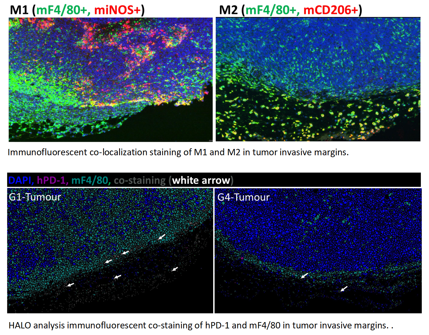
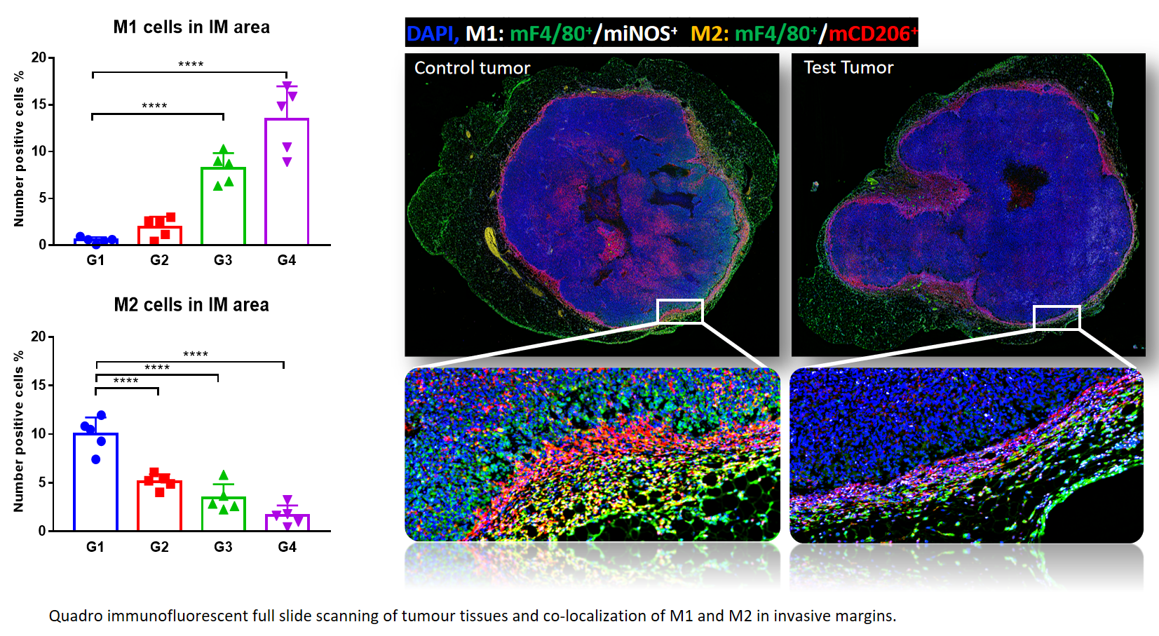
An increase of M1 was found in invasive margin of MC38 tumors, whereas M2 declined significantly in response to pembrolizumab treatment, with a dose-dependent manner. This indicates M1 infiltration facilitated anti-PD-1 mediated tumor growth inhibition.
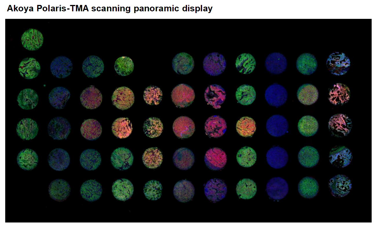
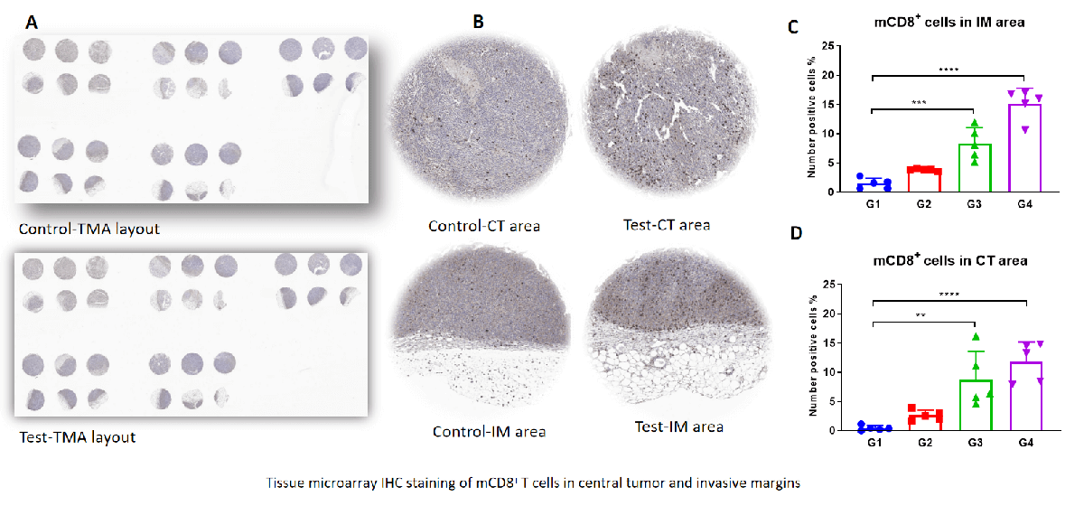
TMA staining shows that CD8+ T (CTL) cells were increased dramatically in both tumor invasive margins and central tumor areas of Test. This is associated with an increased M1/M2 ratio.
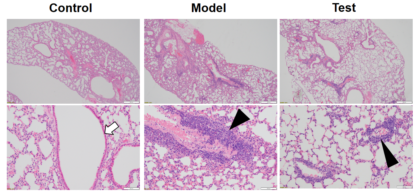
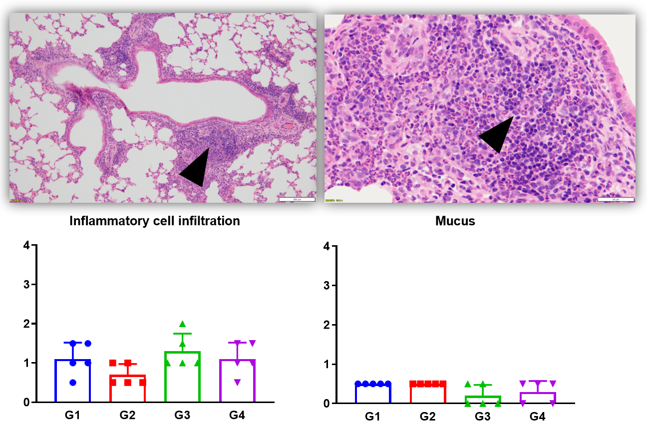
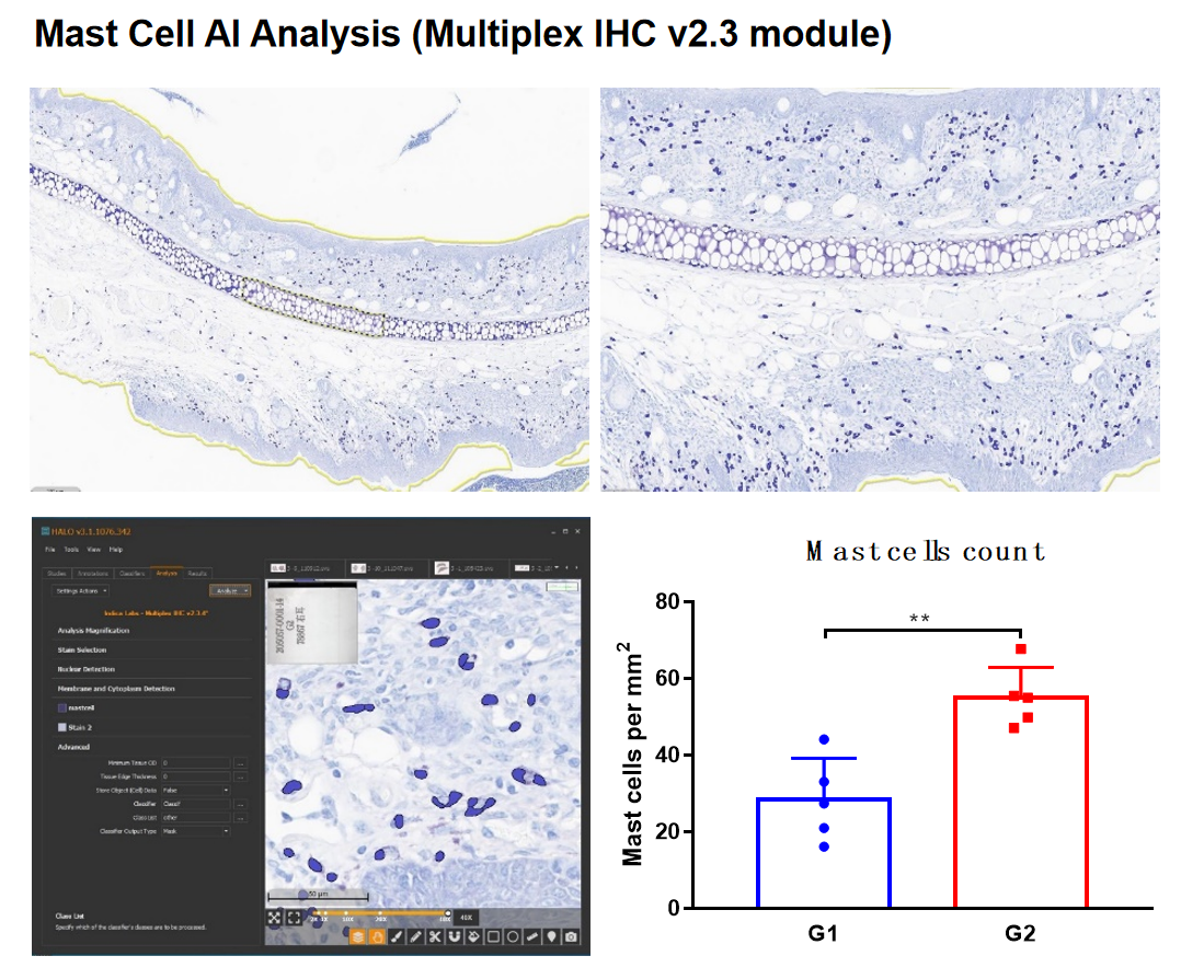
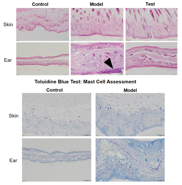
Main features of Asthma:
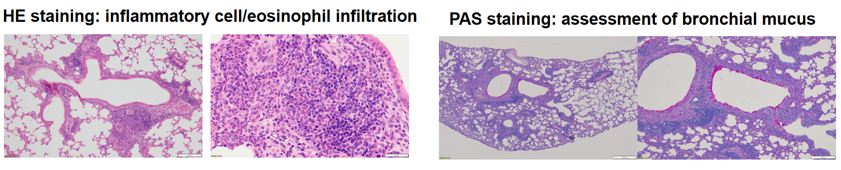
Pathological analysis available:
HE staining- inflammation in psoriasis skin tissue
HE staining- inflammation in atopic dermatitis skin tissue
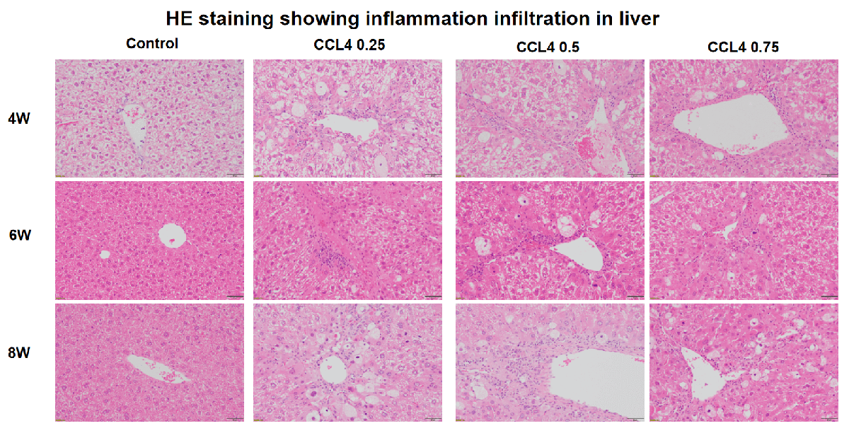
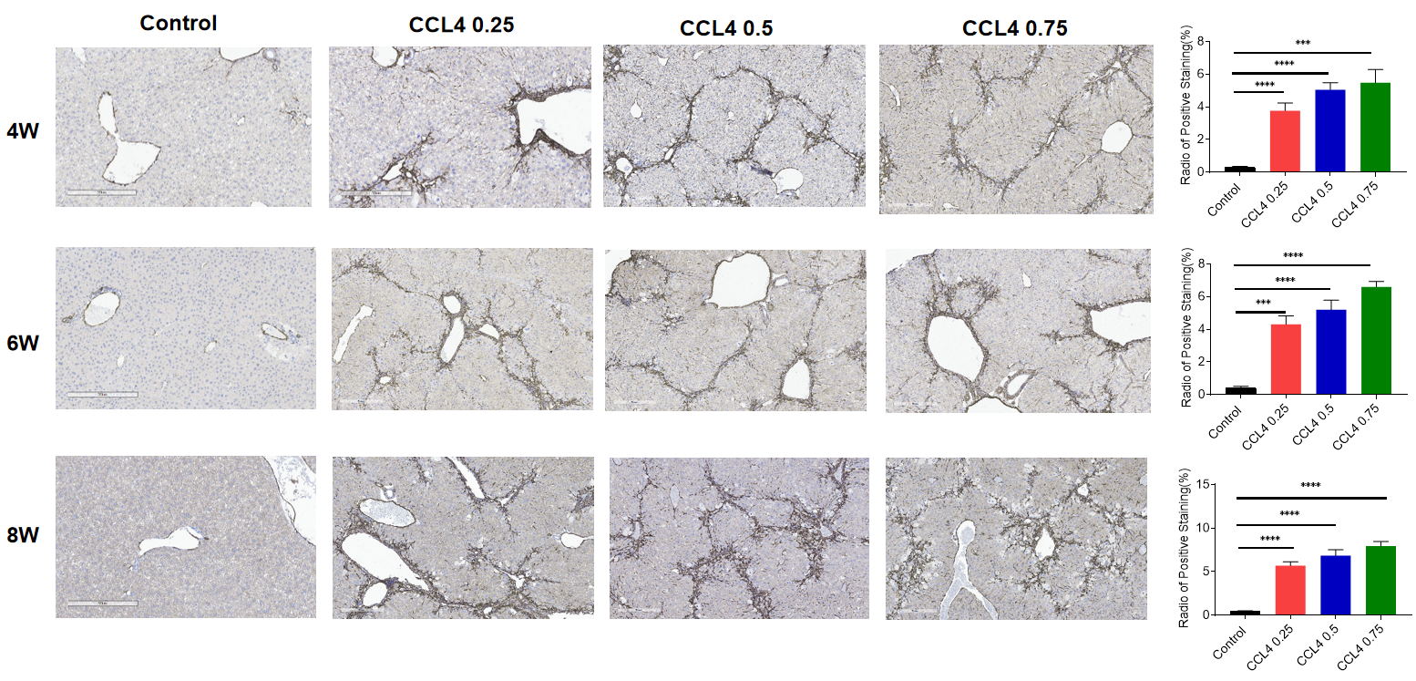
Main features of Non-alcoholic steatohepatitis (NASH):
NASH is a form of NAFLD in which has hepatitis inflammation of the liver and liver cell damage, in addition to fat in the liver. Inflammation and liver cell damage can cause fibrosis, or scarring of the liver. NASH may also lead to cirrhosis or liver cancer.
Immunohistochemistry showing fibroblast marker α-SMA in liver
Main features of IBD:
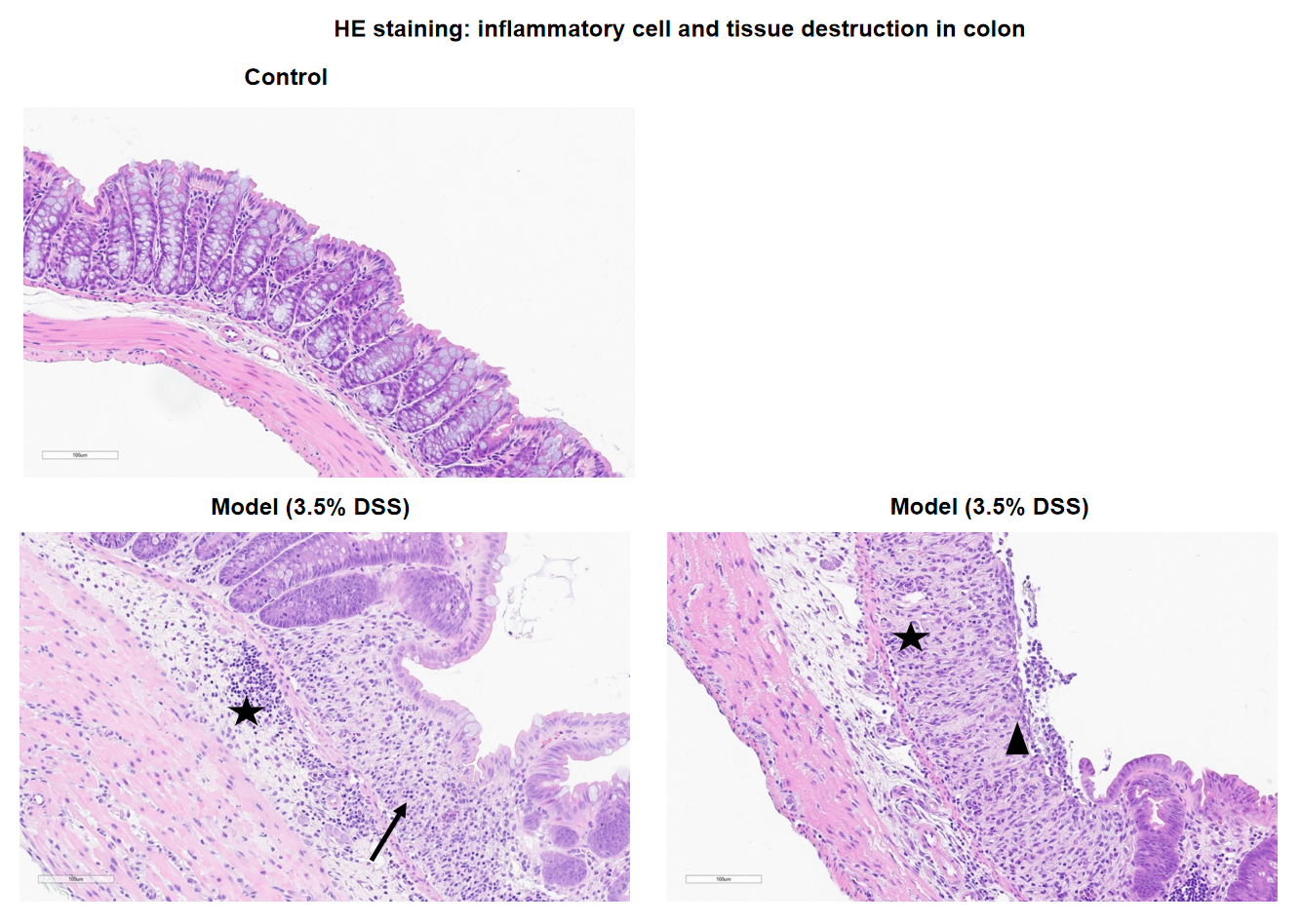
Note: No significant pathological changes were observed in the colon of control group animals microscopically. In the DSS-induced model group, the colon showed crypt loss, ulcer formation, and infiltration of mixed inflammatory cells. Black arrows indicate crypt loss, black triangles indicate ulcer formation in the mucosal layer, and black stars indicate inflammatory cell infiltration.
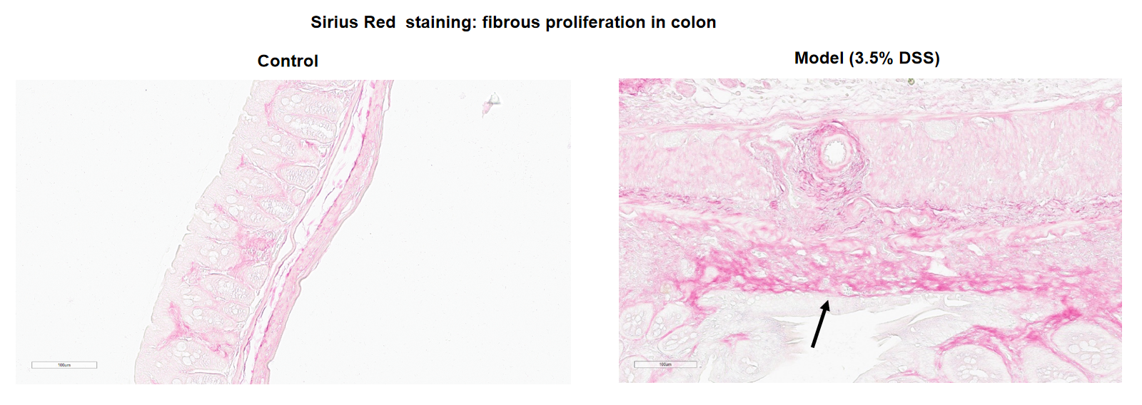
Note: No significant pathological changes were observed in the colon of control group animals microscopically. In the DSS-induced model group, fibrous proliferation was observed in the colon. Black arrows indicate fibrous proliferation.
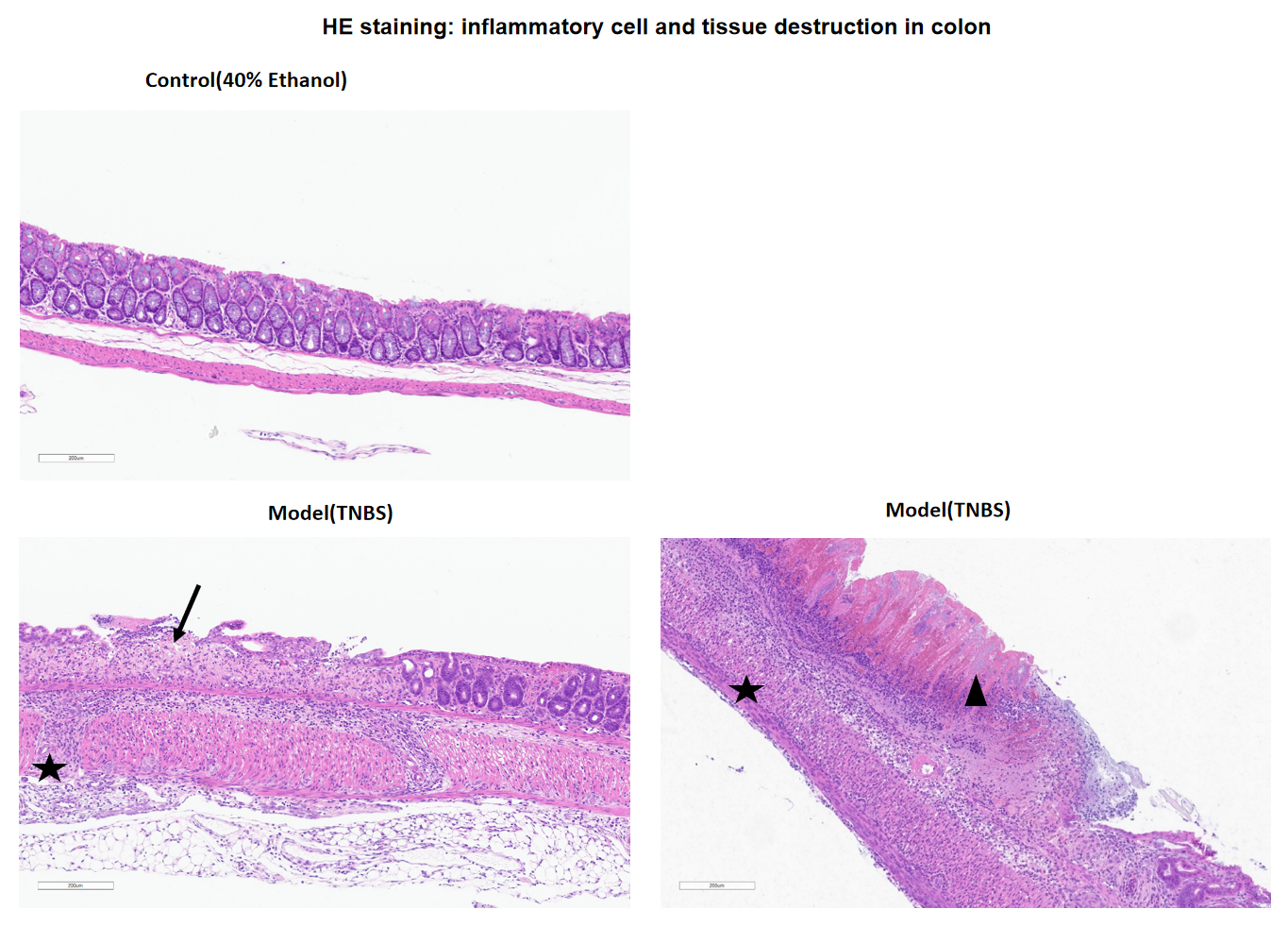
Note: No significant pathological changes were observed in the colon of control group animals microscopically following 40% ethanol instillation. In the TNBS-induced model group, ulcer formation, transmural inflammation, and intestinal necrosis were observed in the colon. Black arrows indicate ulcer formation in the mucosal layer, black triangles indicate intestinal necrosis, and black stars indicate transmural inflammation.
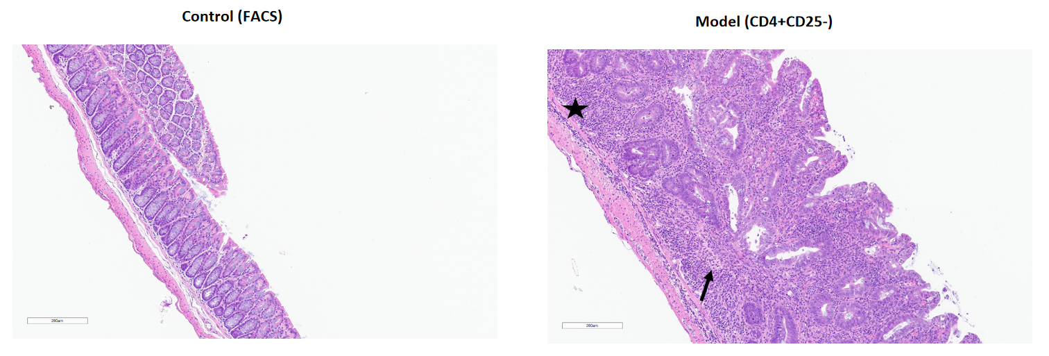
Note: No significant pathological changes were observed in the colon of FACS control group animals microscopically. In the CD4+CD25– adoptive transfer model group, crypt loss and inflammatory cell infiltration were observed in the colon. Black arrows indicate crypt loss, and black stars indicate inflammatory cell infiltration.
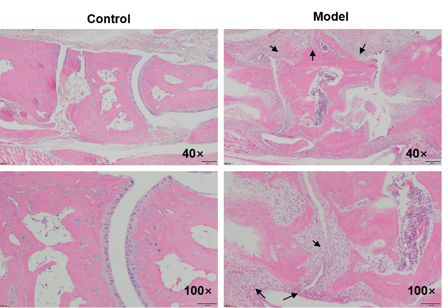
Main features of CIA:
CIA is the most widely studied and used standard model, among the various animal models of rheumatoid arthritis (RA). The pathological features CIA model shared with RA including a proliferative synovitis with infiltration of polymorphonuclear and mononuclear cells, cartilage destruction, bone resorption, pannus formation and fibrosis.
HE staining: inflammatory cell infiltration, cartilage destruction, bone resorption and pannus formation
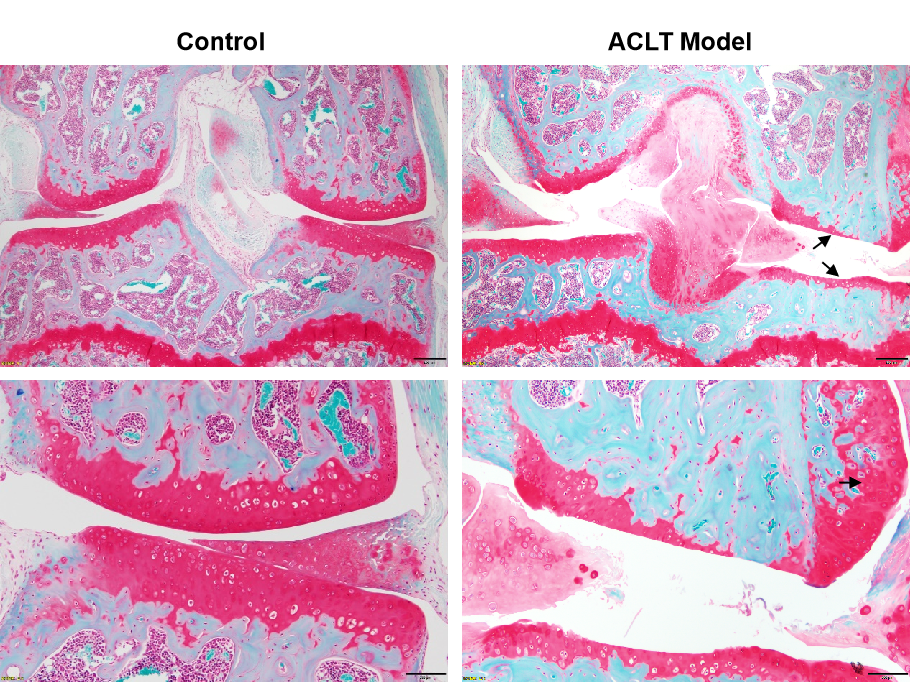
Main features of Surgically-Induced OA Models:
Surgical methods of OA induction work through a combination of joint destabilization, altered joint mechanics, and intra-articular inflammation. Anterior Cruciate Ligament Transection (ACLT )model is induced due to ACL injury, which causes joint destabilization, subsequently leading to Post-traumatic osteoarthritis (PTOA). The ACLT-induced OA model in mouse has many features such as cartilage destruction, osteophyte formation and synovial changes.
Saffron-O and Fast Green staining-cartilage destruction
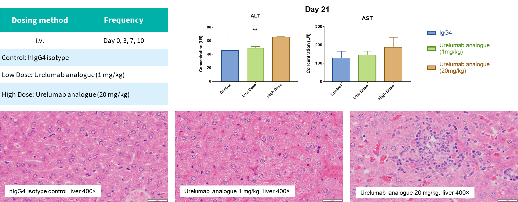
Preclinical toxicology studies are largely focused on drug safety evaluation. Whether a drug is safe and effective is the determining factor in drug development, and toxicity/safety is a major cause for study discontinuation throughout the whole drug development process. The aim is to provide a reliable toxicokinetic basis for human clinical trials by studying the relationship between drug exposure and toxic response in animal models, explaining possible target organs of toxicity and toxic responses, and predicting drug safety in humans.
Team Introduction
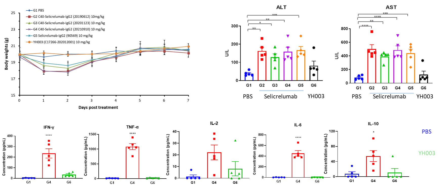
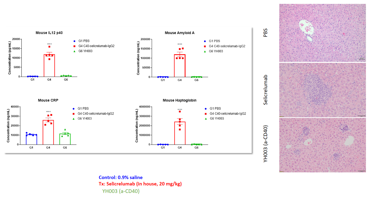
Increased acute phase proteins and liver damage in response to Selicrelumab (analog) in humanized B-hCD40 mice
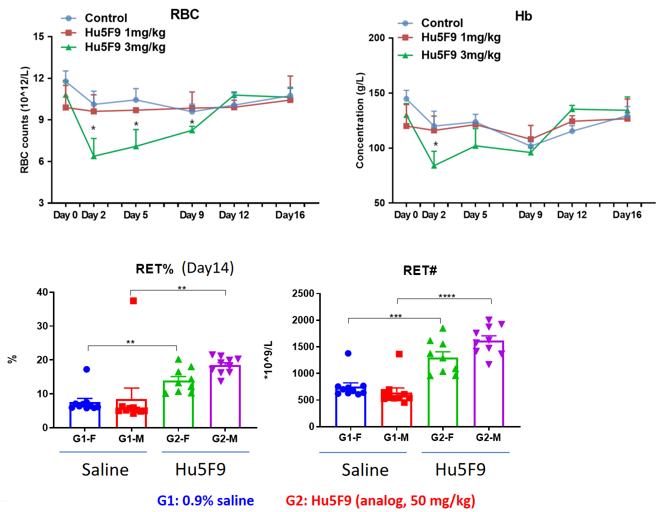
Anaemia and reticulocytosis in humanized B-hSIRPa/hCD47 mice
RBC and Hemoglobin are transiently reduced at the early stage of high dose Hu5F9 treatment and return to normal levels.
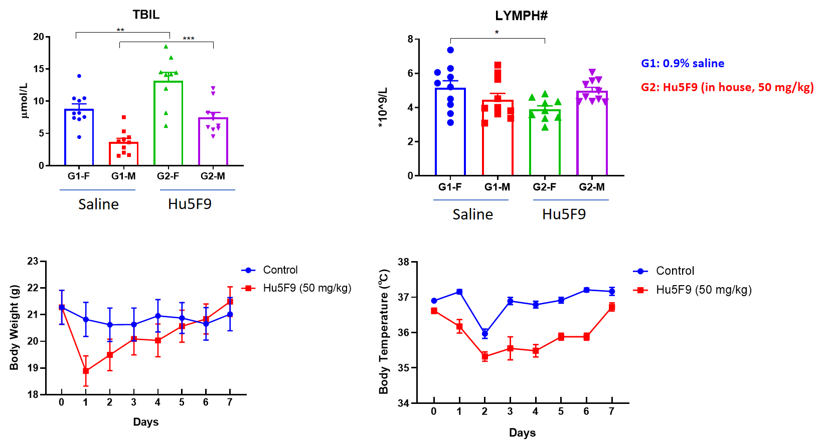
Hyperbilirubinemia, lymphocyte count decreased and chills in humanized B-hSIRPa/hCD47 mice
| Popular Humanized Animal Models For Toxicology Research | ||||
| B-hPD-1 mice | B-h4-1BB mice | B-hPD-1/h4-1BB mice | B-hPD-1/hPD-L1/h4-1BB mice | B-hCCR8 mice |
| B-hLAG3 mice plus | B-hPD-1/hLAG3 mice | B-hCD155 mice | B-hCD6 mice | B-hGLP1R mice |
| B-hCD38 mice | B-hCD147 mice | B-hCD16A mice | B-hCD98HC mice | B-hGCGR mice |
| B-hCD40 mice | B-hCTLA4 mice | B-hGITR mice | B-hTIGIT mice | B-hTSLP/hTSLPR mice |
| B-hCD3E mice | B-hCD3E/hCD38 mice | B-hCD3E/hCD28 mice | B-hCD3E/h4-1BB mice | B-hTNFR2 mice |
| B-hCD3EDG mice | B-hFcRn mice | B-hSIRPA/hCD47 mice | B-hGARP mice | B-hTFR1 mice |
| B-hIL4/hIL4RA mice | B-hIL17A/hIL17F mice | B-hIL36R mice | B-hTNFA mice | B-hRANKL mice |
| B-hIL2RA mice | B-hIL2RB/IL2RG mice | B-hIL13 mice | B-hIL1RAcP mice | B-hIL10RA/IL2RG mice |
