
Biocytogen has developed various animal models, including the APAP-induced and concanavalin A (Con A)-induced acute liver injury models, to investigate disease mechanisms and evaluate new therapeutic strategies for mitigating liver damage and inflammation.
on this page
Acute liver injury (ALI) and autoimmune liver diseases, such as autoimmune hepatitis (AIH), are serious conditions that can lead to liver failure if not properly managed. ALI is typically caused by factors such as toxins, viral infections, or ischemia, leading to liver cell death and inflammation. Autoimmune liver diseases, including AIH, occur when the immune system mistakenly attacks the liver, causing chronic inflammation and potential progression to cirrhosis.
To study these conditions and evaluate potential treatments, Biocytogen has developed various animal models, including the APAP-induced and concanavalin A (Con A)-induced acute liver injury model. These models are crucial for investigating disease mechanisms and testing new therapeutic strategies to mitigate liver damage and inflammation.
APAP is taken up in the intestine within the first 2 hours after oral administration, metabolized in the liver via glucoronidation and sulfonation, and excreted in the urine. A small fraction (10-15%) is metabolized in hepatocytes by cytochrome P450 isomers to the alkylated, highly toxic metabolite N-acetyl-p-benzoquinoneimine (NAPQI). Antioxidant glutathione (GSH) converts NAPQI to the less harmful reduced form, which is then excreted via bile. When glutathione is depleted, increasing amounts of NAPQI bind to mitochondrial proteins and leading to hepatocyte necrosis.

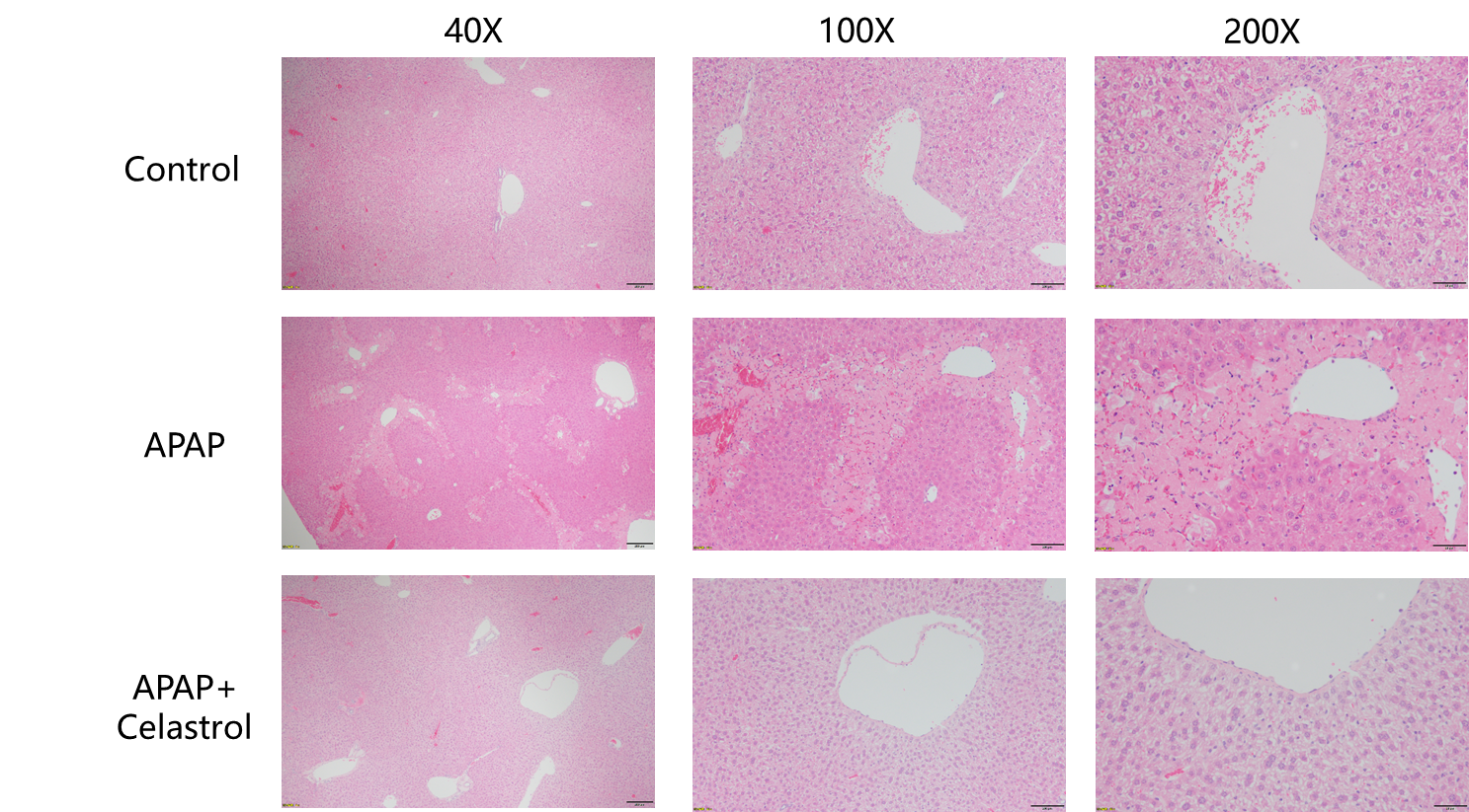
Protective effects of Celastrol in liver injury. Coagulative necrosis and bruising of cells in the marginal zone of the hepatic lobules were seen to varying degrees in the livers of the modeled animals, and the Celastrol treatment group showed significant improvement in the pathology.
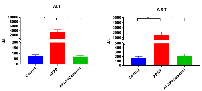
Celastrol treatment alleviate increased ALT and AST enzyme activity induced by APAP. Values are expressed as mean ± SEM. N = 8 mice per group. *p<0.05.
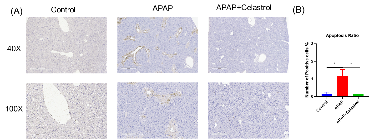
Celastrol reduced apoptosis in APAP induced liver injury. A,Tunel staining showing apoptosis in hepatic cells. B, Statistic data of Tunel positive staining. Values are expressed as mean ± SEM. N = 8 mice per group. *p<0.05.
The pathogenesis and pathological changes in the ConA-induced mouse hepatitis model, in which liver injury is mediated by T cell and macrophage activation, partially mimic those of human autoimmune hepatitis, and therefore this model has been widely used to study the pathogenesis, pathological changes, and clinical treatment of diseases such as autoimmune hepatitis. Hepatocyte apoptosis, necrosis, and leukocyte infiltration are seen as prominent features of immune hepatitis. In this model, activated T cells and macrophages infiltrate the liver tissue mesenchyme and induce the secretion of several pro-inflammatory factors, such as TNF-α, IFN-γ, IL-1β and IL-2.

A, Celastrol treatment alleviate increased ALT and AST enzyme activity induced by ConA . B, Analysis of H&E staining in the protective effect of Celastrol on ConA induced NASH mouse models. Values are expressed as mean ± SEM. N = 8 mice per group. *p<0.05,**p<0.01.
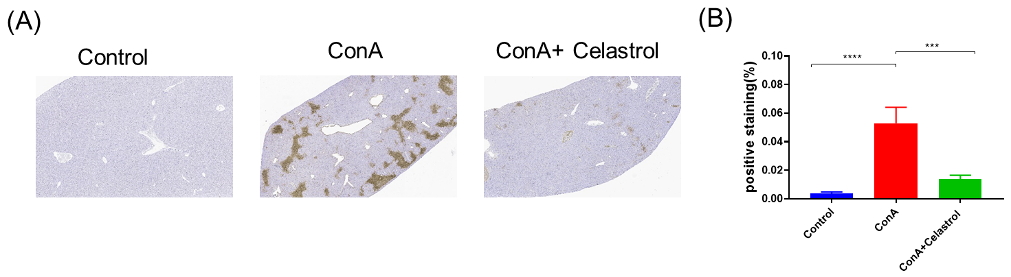
Pathological analysis in the protective effect of Celastrol on ConA induced acute liver injury Mouse Model. A,Tunel staining showing apoptosis in hepatic cells. B, Statistic data of Tunel positive staining. Values are expressed as mean ± SEM. N = 8 mice per group. ***p<0.001.
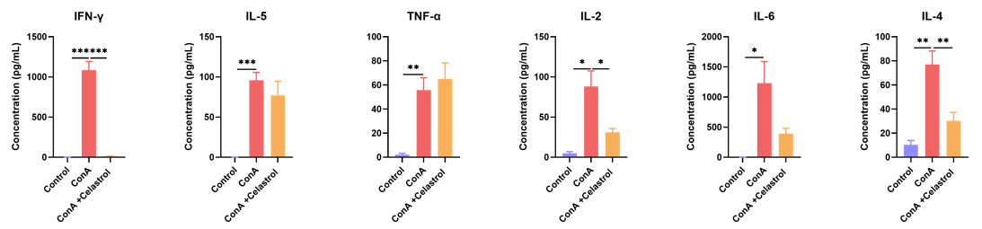
Celastrol treatment alleviate increased cytokines levels induced by Con A.N = 8 mice per group. *p<0.05.
