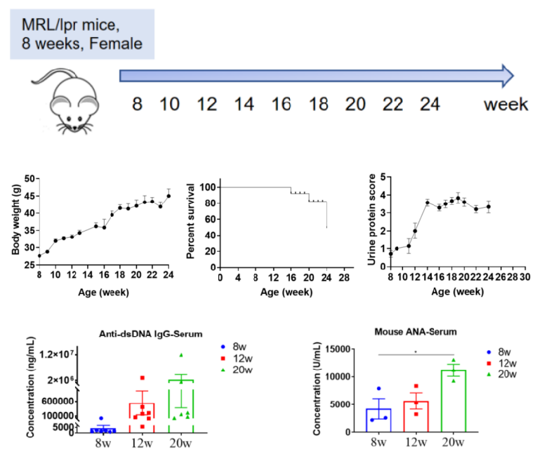
Establishment of Spontaneous SLE Mouse Model in MRL/lpr Mice
| Sample |
Evaluation Index |
| Urine |
Urine protein |
| Serum |
Anti-dsDNA antibody, ANA |
| Pathological detection |
H&E staining |
| IHC staining |
|
Body weight, Survival curve |
Establishment of spontaneous SLE mouse model in MRL/lpr mice. Changes of body weight, urine protein, survival curve, anti-dsDNA and ANA in serum at different time points of MRL/lpr mice.
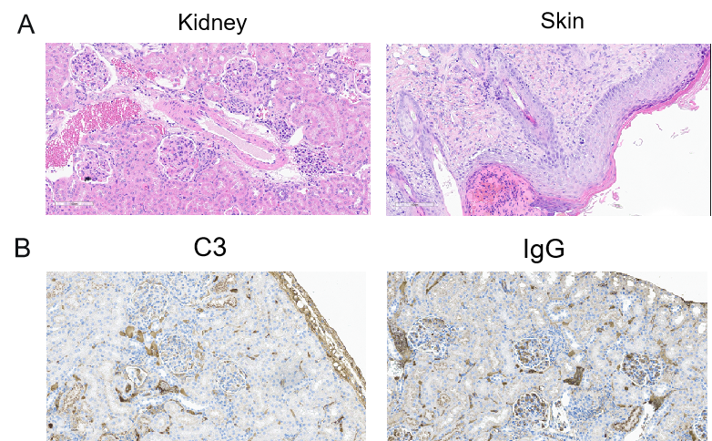
Histological Assessment of Spontaneous SLE in MRL/lpr Mice
Histological analysis of the kidneys of SLE model. A. Glomerular hypertrophy, glomerular cell hyperplasia, and increased matrix were observed in kidney; Epidermal cell hyperplasia, epidermal thickening, hyperkeratosis with hypokeratosis, dermal inflammatory cell infiltration were observed in skin (mice aged 20 weeks). B. C3 and IgG deposition in the kidney of mice aged 20 weeks