Alopecia Areata Model Introduction
Alopecia areata (AA) is an autoimmune disorder characterized by non-scarring hair loss, resulting from T cell-mediated attacks on hair follicles. To investigate the underlying mechanisms and evaluate potential treatments, various alopecia areata mouse models have been developed.
Imiquimod, a topical immune response modifier, activates Toll-like receptor 7 (TLR7), leading to a Th1-skewed immune response characterized by elevated levels of interferon-gamma (IFN-γ) and other pro-inflammatory cytokines. Topical application of imiquimod on mouse models, such as C3H/HeJ and C57BL/6 strains, induces AA-like symptoms, including localized hair loss and immune cell infiltration around hair follicles. This provides a powerful in vivo tool for preclinical studies of AA pathogenesis and therapeutic evaluation.
Establishment of Imiquimod Induced Alopecia Areata Models
- Animals: C3H/HeJ mice, Male, 6-8 weeks
- Reagents: Imiquimod
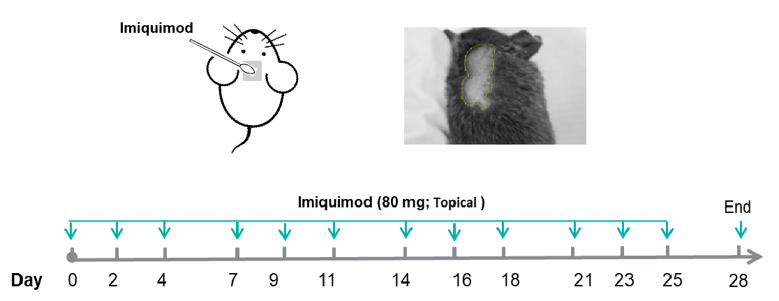
- Methods: Mice receive topical application of imiquimod (80 mg) on neck skin (1.5 cm x 1.5 cm). Image J software is used to calculate the area of hair loss. At the endpoint, spleen and lymph nodes (lymphoglandulae auriculares posteriores) are collected and weighed, and neck skin is collected for H&E staining.
| Readouts |
| Included tests |
Clinical scores |
Alopecia areata area |
| Percentage of alopecia areata |
| Optional tests |
Molecular level |
Protein level (Elisa or Luminex) |
| Spleen, lymphoglandulae auriculares posteriores (LAP) |
Weight |
| Pathological test |
Immune cell infiltration |
| Epidermal thickness |
| Hair follicle number |
| IHC: CD4+, CD8+, MHC I |
Imiquimod Induced Alopecia Areata in C3H/HeJ and C57BL/6 Mice
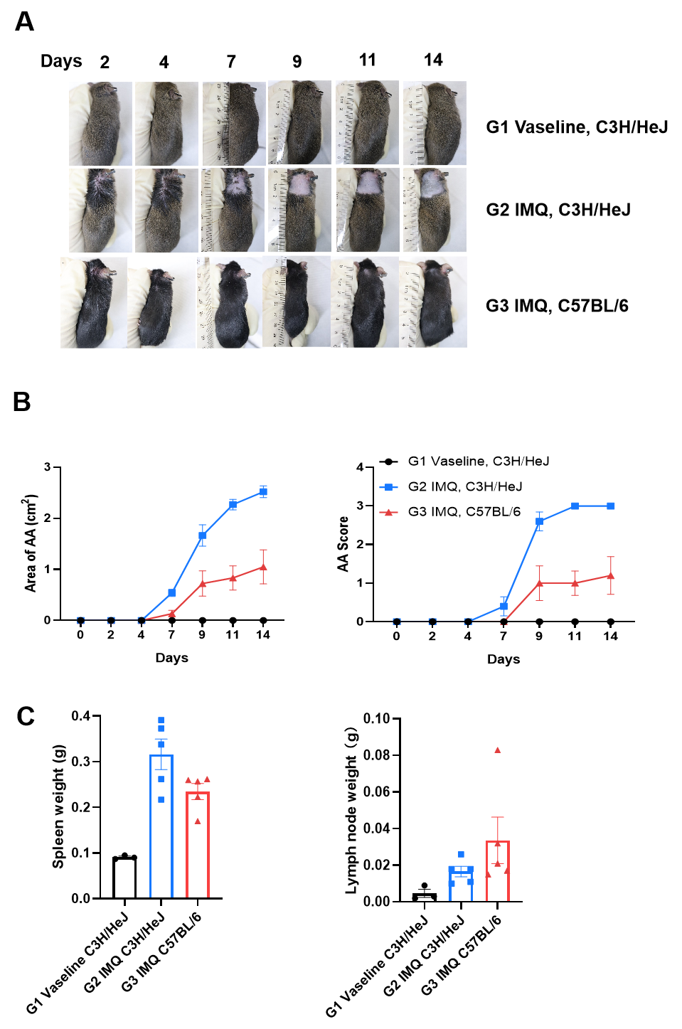
Imiquimod-induced progressive hair loss and systemic immune activation in C3H/HeJ and C57BL/6 Mice. (A) Mice receive topical application of imiquimod (80 mg) on day 0, 2, 4, 7,9,11. Pictures of the hair loss were recorded on day 2, 4, 7, 9, 11 and 14. (B) Image J software were used to calculated the area of hair loss. (C) At the endpoint, spleen and lymphoglandulae auriculares posteriores were collected and weighed. AA: Alopecia Areata, IMQ: Imiquimod.
Pathological Changes in Imiquimod Induced Alopecia Areata Model
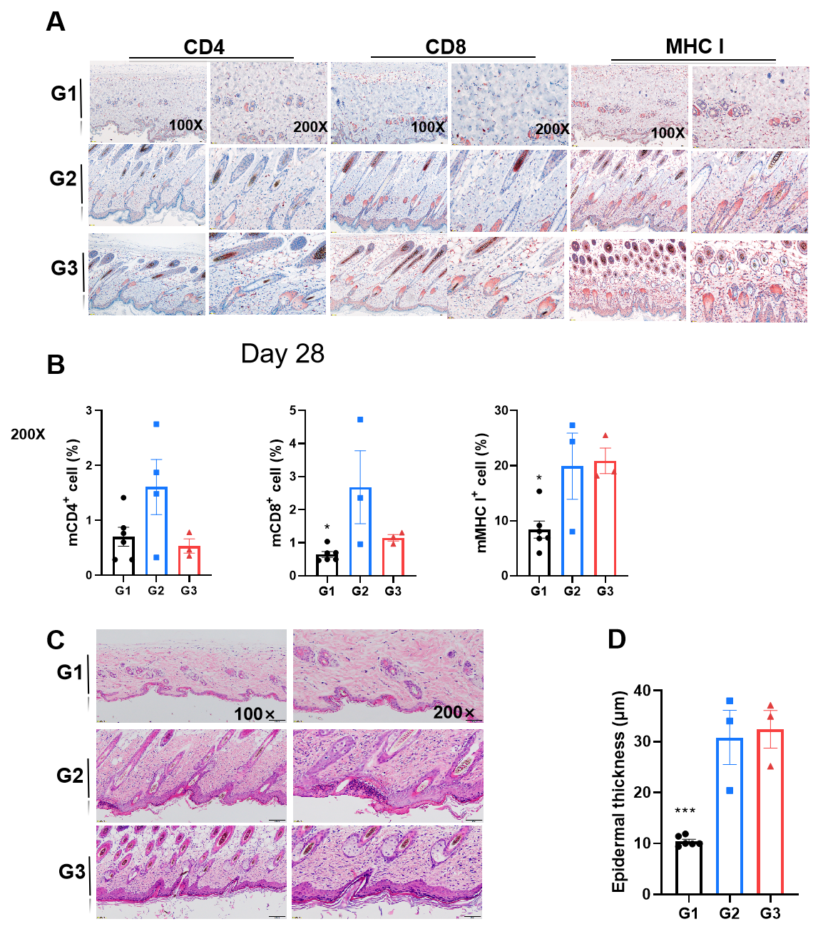
Pathological and immune cell changes in imiquimod-induced alopecia areata mouse model. Neck skin was collected for IHC (A, B) and H&E staining (C, D). In imiquimod induced alopecia areata model mice, observed an increase in CD4+, CD8+ and MHC I+ cells in skin (B). Epidermal was thicker in modeling mice (C, D).