

• 310708
| Product name | B-hEPCAM MC38 |
|---|---|
| Catalog number | 310708 |
| Strain background | C57BL/6 |
| Aliases | DIAR5, EGP-2, EGP40, ESA, HNPCC8, TROP1 |
| Tissue | Colon |
| Disease | Colon carcinoma |
| Species | Mouse |
| Application | B-hEPCAM MC38 cells have the capability to establish tumors in vivo and can be used for efficacy studies. |
on this page
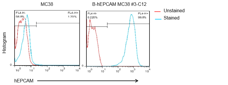
The mouse Epcam gene was replaced by human EPCAM coding sequence in B-hEPCAM MC38 cells. Human EPCAM is highly expressed on the surface of B-hEPCAM MC38 cells.
Gene targeting strategy for B-hEPCAM MC38 cells. The exogenous promoter and human EPCAM coding sequence were inserted to replace part of murine exon 4 and all of exons 5~7. The insertion disrupts the endogenous murine Epcam gene, resulting in a non-functional transcript.
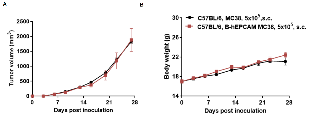
Subcutaneous homograft tumor growth of B-hEPCAM MC38 cells. B-hEPCAM MC38 cells (5x10⁵) and wild-type MC38 cells (5x10⁵) were subcutaneously implanted into C57BL/6 mice (female, 5-8-week-old, n=5). Tumor volume and body weight were measured twice a week. (A) Average tumor volume ± SEM. (B) Body weight (Mean± SEM). Volume was expressed in mm3 using the formula: V=0.5 X long diameter X short diameter2. As shown in panel A, B-hEPCAM MC38 cells were able to establish tumors in vivo and can be used for efficacy studies.
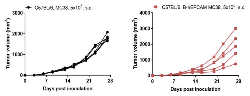
B-hEPCAM MC38 tumor growth of individual mice. B-hEPCAM MC38 cells (5x10⁵) and wild-type MC38 cells (5x10⁵) were subcutaneously implanted into C57BL/6 mice (female, 5-8-week-old, n=5). As shown in panel, B-hEPCAM MC38 cells were able to establish tumors in vivo and can be used for efficacy studies.
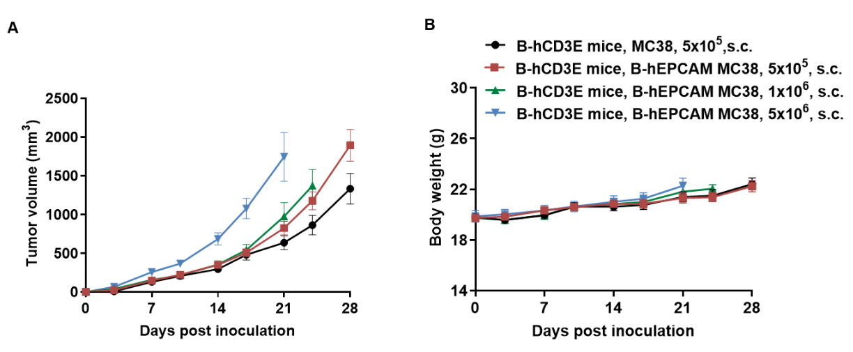
Subcutaneous homograft tumor growth of B-hEPCAM MC38 cells. B-hEPCAM MC38 cells (5x10⁵, 1x10⁶, 5x10⁶) and wild-type MC38 cells (5x10⁵) were subcutaneously implanted into B-hCD3E mice (female, 8-week-old, n=6). Tumor volume and body weight were measured twice a week. (A) Average tumor volume ± SEM. (B) Body weight (Mean± SEM). Volume was expressed in mm3 using the formula: V=0.5 X long diameter X short diameter2. As shown in panel A, B-hEPCAM MC38 cells were able to establish tumors in vivo and can be used for efficacy studies.

B-hEPCAM MC38 tumor growth of individual mice. B-hEPCAM MC38 cells (5x10⁵, 1x10⁶, 5x10⁶) and wild-type MC38 cells (5x10⁵) were subcutaneously implanted into B-hCD3E mice (female, 8-week-old, n=6). As shown in panel, B-hEPCAM MC38 cells were able to establish tumors in vivo and can be used for efficacy studies.
