

• 322242
| Product name | B-hFOLR1 MC38 |
|---|---|
| Catalog number | 322242 |
| Strain background | C57BL/6 |
| Aliases | FBP, FOLR, NCFTD, Fralpha |
| Tissue | Colon |
| Disease | Colon carcinoma |
| Species | Mouse |
| Application | B-hFOLR1 MC38 |
on this page
Origin: The MC38 cell line is derived from C57BL/6 murine colon adenocarcinoma cells. The cell line is a commonly used murine model for colorectal carcinoma.
Background Information: The protein encoded by this gene is a member of the folate receptor family. Members of this gene family bind folic acid and its reduced derivatives, and transport 5-methyltetrahydrofolate into cells. This gene product is a secreted protein that either anchors to membranes via a glycosyl-phosphatidylinositol linkage or exists in a soluble form. Mutations in this gene have been associated with neurodegeneration due to cerebral folate transport deficiency.
Gene targeting strategy: The human FOLR1 coding sequence was inserted into the exon 4 of mouse Folr1 in B-hFOLR1 MC38 cells. Human FOLR1 is highly expressed on the surface of B-hFOLR1 MC38 cells.
Application: B-hFOLR1 MC38 cells have the capability to establish tumors in vivo and can be used for efficacy studies.
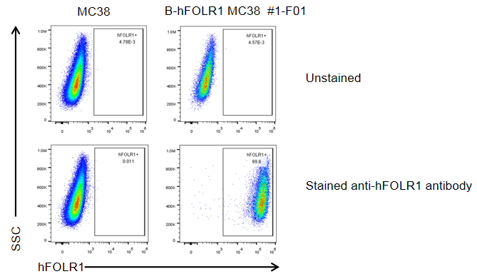
FOLR1 expression analysis in B-hFOLR1 MC38 cells by flow cytometry. Single cell suspensions from wild-type MC38 and B-hFOLR1 MC38 cultures were stained with species-specific anti-FOLR1 antibody(Biolegend, 908304). Human FOLR1 was detected on the surface of B-hFOLR1 MC38 cells but not wild-type MC38 cells. The 1-F01 clone of B-hFOLR1 MC38 cells was used for in vivo experiments.
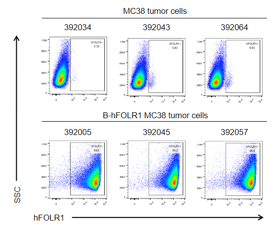
B-hFOLR1 MC38 cells were subcutaneously transplanted into C57BL/6 mice (n=8 or 6), and on 31 days post inoculation, tumor cells were harvested and assessed for human FOLR1 expression by flow cytometry. As shown, human FOLR1 was highly expressed on the surface of tumor cells. Therefore, B-hFOLR1 MC38 cells can be used for in vivo efficacy studies of novel FOLR1 therapeutics.
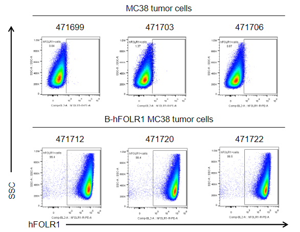
B-hFOLR1 MC38 cells were subcutaneously transplanted into B-hFOLR1/hFOLR2 mice (n=6), and on 31 days post inoculation, tumor cells were harvested and assessed for human FOLR1 expression by flow cytometry. As shown, human FOLR1 was highly expressed on the surface of tumor cells. Therefore, B-hFOLR1 MC38 cells can be used for in vivo efficacy studies of novel FOLR1 therapeutics.
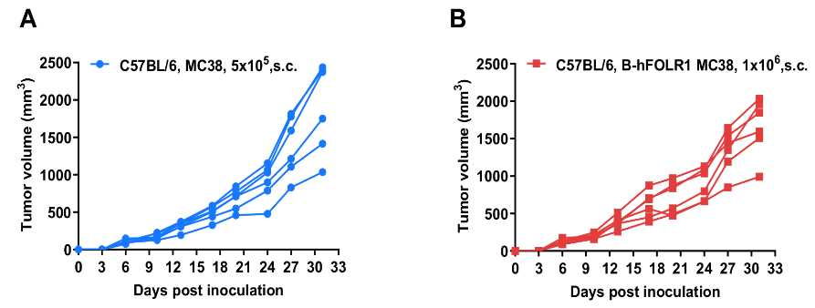
B-hFOLR1 MC38 tumor cells growth of individual mice. B-hFOLR1 MC38 cells (1x10⁶) and wild-type MC38 cells (5x10⁵) were subcutaneously implanted into C57BL/6 mice (female, 8-week-old, n=8 or 6). As shown in panel, B-hFOLR1 MC38 cells were able to establish tumors in vivo and can be used for efficacy studies.
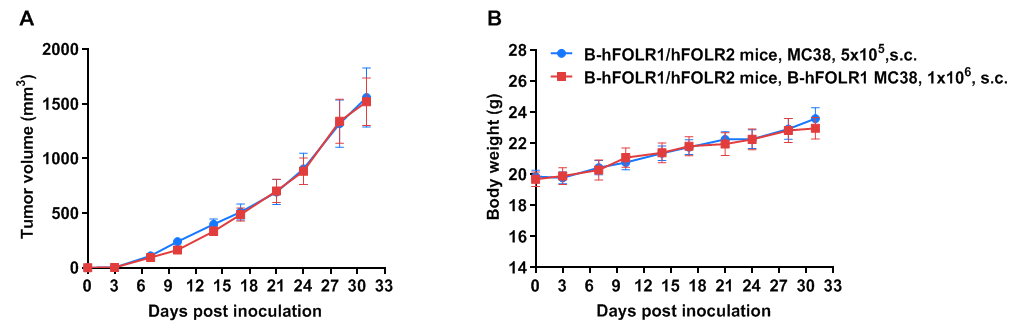
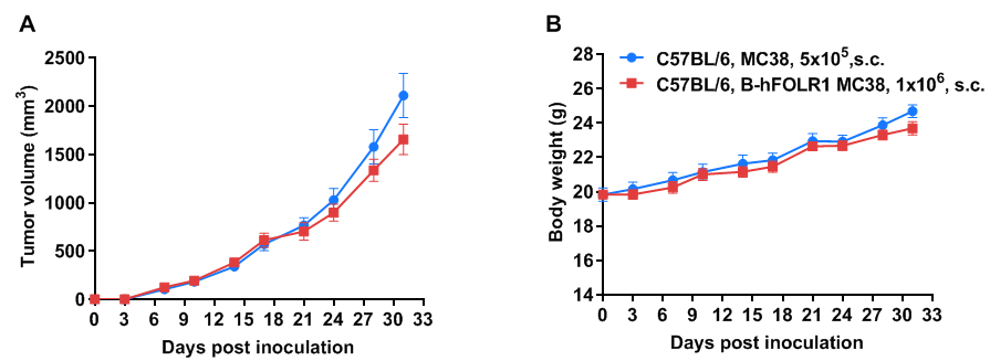
Subcutaneous homograft tumor growth of B-hFOLR1 MC38 cells. B-hFOLR1 MC38 cells (1x10⁶) and wild-type MC38 cells (5x10⁵) were subcutaneously implanted into B-hFOLR1/hFOLR2 mice (female, 8-week-old, n=7). Tumor volume and body weight were measured twice a week. (A) Average tumor volume ± SEM. (B) Body weight (Mean± SEM). Volume was expressed in mm3 using the formula: V=0.5 X long diameter X short diameter2. As shown in panel A, B-hFOLR1 MC38 cells were able to establish tumors in vivo and can be used for efficacy studies.
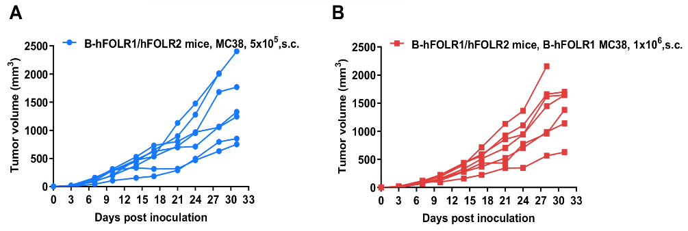
B-hFOLR1 MC38 tumor cells growth of individual mice. B-hFOLR1 MC38 cells (1x10⁶) and wild-type MC38 cells (5x10⁵) were subcutaneously implanted into B-hFOLR1/hFOLR2 mice (female, 8-week-old, n=6). As shown in panel, B-hFOLR1 MC38 cells were able to establish tumors in vivo and can be used for efficacy studies.
