

• 322404
| Product name | B-HLA-A2.1/hMAGEA4 MC38 |
|---|---|
| Catalog number | 322404 |
| Strain background | C57BL/6 |
| Aliases | IMD43, AMYLD6, MHC1D4; HLAA; CT1.4, MAGE4, MAGE4A, MAGE4B, MAGE-41, MAGE-X2 |
| Tissue | Colon |
| Disease | Colon carcinoma |
| Species | Mouse |
| Application | B-HLA-A2.1/hMAGEA4 MC38 tumor models can be used for preclinical evaluation of cancer vaccines. |
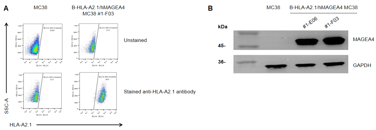
HLA-A2.1 and MAGEA4 expression analysis in B-HLA-A2.1/hMAGEA4 MC38 cells by flow cytometry, respectively. Single cell suspensions from wild-type MC38 and B-HLA-A2.1/hMAGEA4 MC38 #1-F03 cultures were detected with species-specific anti-HLA-A2.1 antibody (Biolegend, 343306) and anti-human MAGEA4 (Invitrogen, 35-6300), respectively. Human HLA-A2.1 was detected on the surface of B-HLA-A2.1/hMAGEA4 MC38 cells but not wild-type MC38 cells(A). Human MAGEA4 was detected in the B-HLA-A2.1/hMAGEA4 MC38 cells but not wild-type MC38 cells(B).
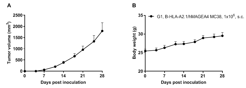
Subcutaneous tumor growth of B-HLA-A2.1/hMAGEA4 MC38 cells. B-HLA-A2.1/hMAGEA4 MC38 cells (1×10⁶) were subcutaneously implanted into B-HLA-A2.1 mice (male, 7-week-old, n=6). Tumor volume and body weight were measured twice a week. (A) Average tumor volume. (B) Body weight. Volume was expressed in mm3 using the formula: V=0.5 × long diameter × short diameter2. Results indicate that B-HLA-A2.1/hMAGEA4 MC38 cells were able to establish tumors in vivo and can be used for efficacy studies. Values are expressed as mean ± SEM.
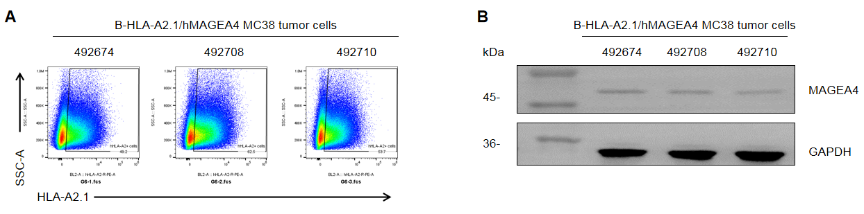
HLA-A2.1 and MAGEA4 expression evaluated in B-HLA-A2.1/hMAGEA4 MC38 tumor cells by flow cytometry, respectively. B-HLA-A2.1/hMAGEA4 MC38 cells were subcutaneously transplanted into B-HLA-A2.1 mice (male, 7-week-old, n=6). Upon conclusion of the experiment, tumor cells were harvested and assessed with species-specific anti-HLA-A2.1 antibody (Biolegend, 343306) and anti-human MAGEA4 (Invitrogen, 35-6300), respectively. Human HLA-A2.1 was highly expressed on the surface of tumor cells(A). Human MAGEA4 was detected in the tumor cells(B). Therefore, B-HLA-A2.1/hMAGEA4 MC38 cells can be used for in vivo efficacy studies evaluating cancer vaccines.
