

• 310687
| Product name | B-hPD-L1 B16-F10 |
|---|---|
| Catalog number | 310687 |
| Strain background | C57BL/6 |
| Aliases | CD274, PDCD1L1 |
| Tissue | Skin |
| Disease | Melanoma |
| Species | Mouse |
| Application | B-hPD-L1 B16-F10 |
on this page
The mouse Pdl1 gene was replaced by human PD-L1 coding sequence in B-hPD-L1 B16-F10 cells. Human PD-L1 is highly expressed on the surface of B-hPD-L1 B16-F10 cells.
The exogenous promoter and human PD-L1 coding sequence was inserted to replace part of murine exon 3. The insertion disrupts the endogenous murine Pdl1 gene, resulting in a non-functional transcript.
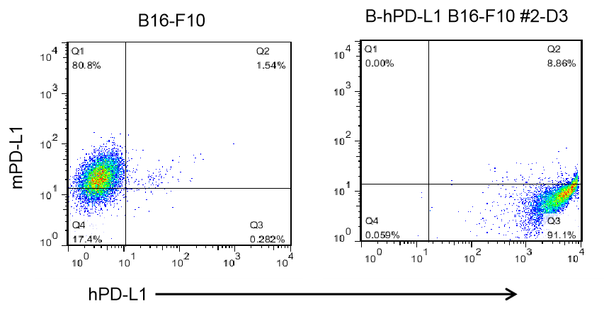
PD-L1 expression analysis in B-hPD-L1 B16-F10 cells by flow cytometry. Single cell suspensions from wild-type B16-F10 and B-hPD-L1 B16-F10 cultures were stained with species-specific anti-PD-L1 antibody. Human PD-L1 was detected on the surface of B-hPD-L1 B16-F10 cells but not wild-type B16-F10 cells. The 2-D3 clone of B-hPD-L1 B16-F10 cells was used for in vivo experiments.
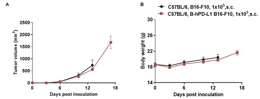
Subcutaneous homograft tumor growth of B-hPD-L1 B16-F10 cells. Wild-type B16-F10 and B-hPD-L1 B16-F10 cells (1x10⁵) were subcutaneously implanted into C57BL/6 mice (female, 5-8-week-old, n=5). Tumor volume and body weight were measured twice a week. (A) Average tumor volume ± SEM. (B) Body weight (Mean± SEM). Volume was expressed in mm3 using the formula: V=0.5 X long diameter X short diameter2. As shown in panel A, B-hPD-L1 B16-F10 cells were able to establish tumors in vivo and can be used for efficacy studies.
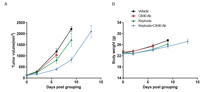
The B-hPD-L1 B16-F10 cell line was subcutaneously implanted into homozygous B-hPD-1/hOX40 mice. Mice were grouped when tumor size reached approximately 150 ± 50 mm3 (n = 5). (A) Mean tumor volume ± SEM; (B) Mean body weight of mice ± SEM. The result shows a moderate antitumor activity by the Keytruda or OX40 Ab compared with the vehicle control group. The data also indicated that Keytruda combined with OX40 Ab potentiated the antitumor activity of the Keytruda or OX40 Ab alone.
