

• 310890
| Product name | B-hPD-L1 low B16-F10 |
|---|---|
| Catalog number | 310890 |
| Strain background | C57BL/6 |
| Aliases | B7-H, B7H1, PDCD1L1, PDCD1LG1 |
| Tissue | Skin |
| Disease | Melanoma |
| Species | Mouse |
| Application | B-hPD-L1 low B16-F10 |
on this page
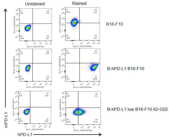
The mouse Pdl1 gene was replaced by human PD-L1 coding sequence in B-hPD-L1 low B16-F10 cells. Human PD-L1 is expressed on the surface of B-hPD-L1 low B16-F10 cells.
Gene targeting strategy for B-hPD-L1 low B16-F10 cells. The exogenous promoter and human PD-L1 coding sequence were inserted to replace part of murine exon 3. The insertion disrupts the endogenous murine Pdl1 gene, resulting in a non-functional transcript.
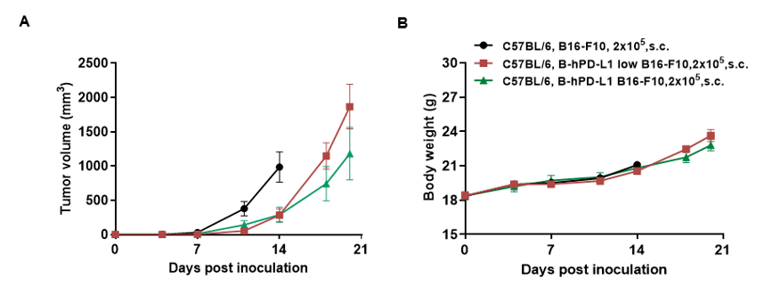
Subcutaneous homograft tumor growth of B-hPD-L1 low B16-F10 cells. Wild-type B16-F10, B-hPD-L1 B16-F10 and B-hPD-L1 low B16-F10 cells (2x10⁵) were subcutaneously implanted into C57BL/6 mice (female, 7-8-week-old, n=5). Tumor volume and body weight were measured twice a week. (A) Average tumor volume ± SEM. (B) Body weight (Mean± SEM). Volume was expressed in mm3 using the formula: V=0.5 X long diameter X short diameter2. As shown in panel A, B-hPD-L1 low B16-F10 cells were able to establish tumors in vivo and can be used for efficacy studies.
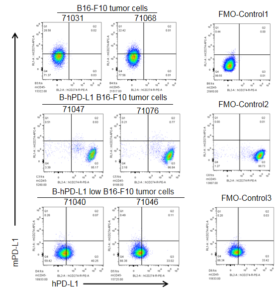
B-hPD-L1 low B16-F10 and B-hPD-L1 B16-F10 cells were subcutaneously transplanted into C57BL/6 mice (n=5). At the end of the experiment, tumor cells were harvested and assessed for human PD-L1 expression by flow cytometry. As shown, human PD-L1 was expressed on the surface of B-hPD-L1 low B16-F10 tumor cells, and it was expressed highly on the surface of B-hPD-L1 B16-F10 tumor cells. Therefore, B-hPD-L1 low B16-F10 cells can be used for in vivo efficacy studies of novel PD-L1 therapeutics.
FMO Control: tumor cells only stained anti-hPD-L1 antibody.
