

• 321917
| Product name | B-hPD-L1 plus/hHER2 MC38 |
|---|---|
| Catalog number | 321917 |
| Strain background | C57BL/6 |
| Aliases | CD274, B7-H, B7H1, PD-L1, PDCD1L1, PDCD1LG1, PDL1, hPD-L1, CD274 molecule,Programmed cell death 1 ligand 1; ERBB2, CD340, HER-2, HER-2/neu, HER2, MLN 19, NEU, NGL, TKR1, VSCN2, c-ERB-2, c-ERB2, erb-b2 receptor tyrosine kinase 2 |
| Tissue | Colon |
| Disease | Colon carcinoma |
| Species | Mouse |
| Application | B-hPD-L1 plus/hHER2 MC38 |
on this page
The mouse Pdl1 gene was replaced by human PD-L1 coding sequence in B-hPD-L1 plus/hHER2 MC38 cells. Human PD-L1 is highly expressed on the surface of B-hPD-L1 plus/hHER2 MC38 cells.
The mouse Erbb2 gene was replaced by human ERBB2 coding sequence in B-hPD-L1 plus/hHER2 MC38 cells. Human ERBB2 is highly expressed on the surface of B-hPD-L1 plus/hHER2 MC38 cells.
Gene targeting strategy for B-hPD-L1 plus/hHER2 MC38 cells.
The exogenous promoter and human PD-L1 coding sequence was inserted to replace part of murine exon 3. The insertion disrupts the endogenous murine Pdl1 gene, resulting in a non-functional transcript.
The exogenous CAG promoter and human ERBB2 coding sequence was inserted to replace part of murine exon 2 and all of exons 3-7. The insertion disrupts the endogenous murine Erbb2 gene, resulting in a non-functional transcript.
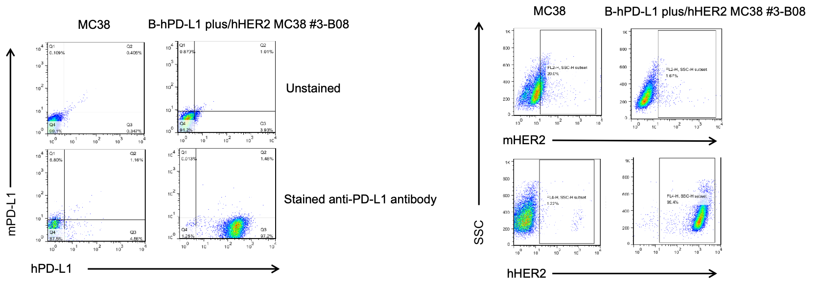
PD-L1 and HER2 expression analysis in B-hPD-L1 plus/hHER2 MC38 cells by flow cytometry. Single cell suspensions from wild-type MC38 and B-hPD-L1 plus/hHER2 MC38 cultures were stained with species-specific anti-PD-L1 and anti-HER2 antibody. Human PD-L1 and HER2 were detected on the surface of B-hPD-L1 plus/hHER2 MC38 cells but not wild-type MC38 cells. The 3-B08 clone of B-hPD-L1 plus/hHER2 MC38 cells was used for in vivo tumor growth assays.
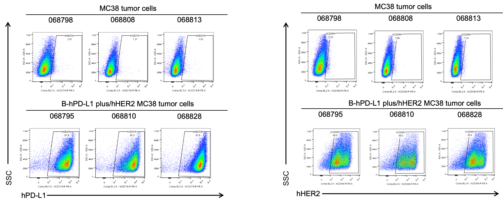
B-hPD-L1 plus/hHER2 MC38 cells were subcutaneously transplanted into C57BL/6 mice (n=6), and on 39 days post inoculation, tumor cells were harvested and assessed for human PD-L1 and HER2 expression by flow cytometry. As shown, human PD-L1 and HER2 were highly expressed on the surface of tumor cells. Therefore, B-hPD-L1 plus/hHER2 MC38 cells can be used for in vivo efficacy studies of PD-L1 and HER2 therapeutics.
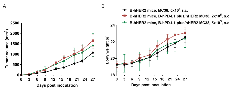
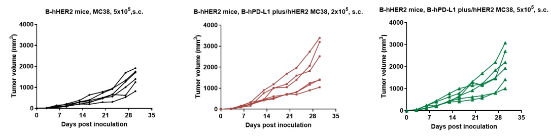

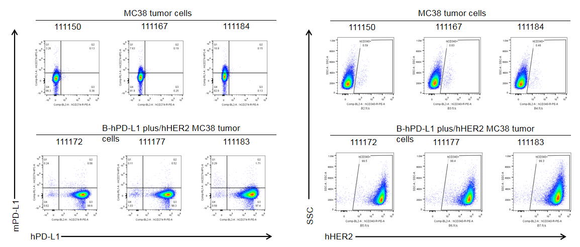
B-hPD-L1 plus/hHER2 MC38 cells were subcutaneously transplanted into homozygous B-hHER2 mice (n=6), and on 30 days post inoculation, tumor cells were harvested and assessed for human PD-L1 and HER2 expression by flow cytometry. As shown, human PD-L1 and HER2 were highly expressed on the surface of tumor cells. Therefore, B-hPD-L1 plus/hHER2 MC38 cells can be used for in vivo efficacy studies of PD-L1 and HER2 therapeutics.
