

• 322214
| Product name | B-hPD-L1 plus/hPCSK9 MC38 |
|---|---|
| Catalog number | 322214 |
| Strain background | C57BL/6 |
| Aliases | CD274, PDCD1L1 FH3, FHCL3, HCHOLA3, LDLCQ1, NARC-1, NARC1, PC9 |
| Tissue | Colon |
| Disease | Colon carcinoma |
| Species | Mouse |
| Application | B-hPD-L1 plus/hPCSK9 MC38 |
on this page
The mouse Pd-l1 gene and Pcsk9 gene were replaced by human PD-L1 and PCSK9 coding sequence in B-hPD-L1 plus/hPCSK9 MC38 cells. Human PD-L1 and human PCSK9 are highly expressed in B-hPD-L1 plus/hPCSK9 MC38.
Gene targeting strategy for B-hPD-L1 plus/hPCSK9 MC38 cells.The exogenous promoter and human PD-L1 coding sequence was inserted to replace partial exon 3 of mouse Pd-l1 gene. The insertion disrupts the endogenous murine Pd-l1 gene, resulting in a non-functional transcript. The exogenous promoter and human PCSK9 coding sequence was inserted to replace part of murine exons 1-3. The insertion disrupts the endogenous murine Pcsk9 gene, resulting in a non-functional transcript.
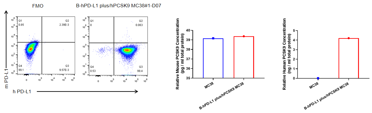
PD-L1 and PCSK9 expression analysis in B-hPD-L1 plus/hPCSK9 MC38 cells. Single cell suspensions from wild-type MC38 and B-hPD-L1 plus/hPCSK9 MC38 cultures were stained with species-specific anti-PD-L1 antibody. Human PD-L1 was detected on the surface of B-hPD-L1 plus/hPCSK9 MC38 cells but not wild-type MC38 cells. Human PCSK9 was detected by ELISA in B-hPD-L1 plus/hPCSK9 MC38 cells but not wild-type MC38 cells. The 1-D07 clone of B-hPD-L1 plus/hPCSK9 MC38 cells was used for in vivo experiments.
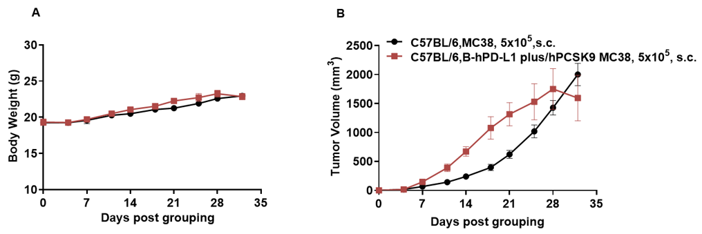
Subcutaneous homograft tumor growth of B-hPD-L1 plus/hPCSK9 MC38 cells. B-hPD-L1 plus/hPCSK9 MC38 cells (5x10⁵) and wild-type MC38 cells (5x10⁵) were subcutaneously implanted into C57BL/6N mice (female, 7-week-old, n=6). Tumor volume and body weight were measured twice a week. (A) Average tumor volume ± SEM. (B) Body weight (Mean± SEM). Volume was expressed in mm3 using the formula: V=0.5 X long diameter X short diameter2. As shown in panel A, B-hPD-L1 plus/hPCSK9 MC38 cells were able to establish tumors in vivo and can be used for efficacy studies.
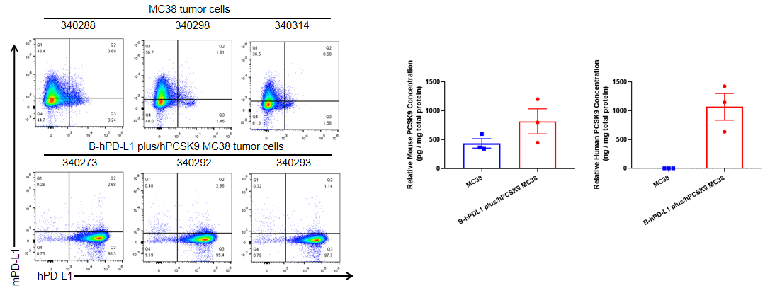
PD-L1 and PCSK9 expression analysis in B-hPD-L1 plus/hPCSK9 MC38 cells. B-hPD-L1 plus/hPCSK9 MC38 cells were subcutaneously transplanted into C57BL/6 mice (n=6), and on 32 days post inoculation, tumor cells were harvested and assessed for human PD-L1 and human PCSK9 expression. As shown, human PD-L1 was highly expressed on the surface of tumor cells and human PCSK9 was secreted in large quantities. Therefore, B-hPD-L1 plus/hPCSK9 MC38 cells can be used for in vivo efficacy studies.
