
• 322265
| Product name | B-Tg(Luc-EGFP) U-87 MG |
|---|---|
| Catalog number | 322265 |
| Tissue | Brain |
| Disease | Glioblastoma |
| Species | Human |
| Application | The B-Tg(Luc-EGFP) U-87 MG cell line stably expresses luciferase, and can be used to evaluate drugs by monitoring the activity of anti-glioblastoma antibodies in xenograft animal models through in vivo bioluminescence imaging. |
on this page
The exogenous promoter and luciferase-EGFP coding sequence were randomly inserted into the U-87 MG cells.
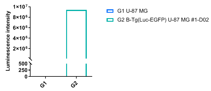
Luminescence signal intensity of B-Tg(Luc-EGFP) U-87 MG cells. Single cell suspensions from wild-type U-87 MG and B-Tg(Luc-EGFP) U-87 MG #1-D02 cultures were measured using the Bright-GloTM luciferase Assay (Promega, Catalog No. E4030). B-Tg(Luc-EGFP) U-87 MG cells have a strong luminescence signal that is not present in wild-type U-87 MG cells.
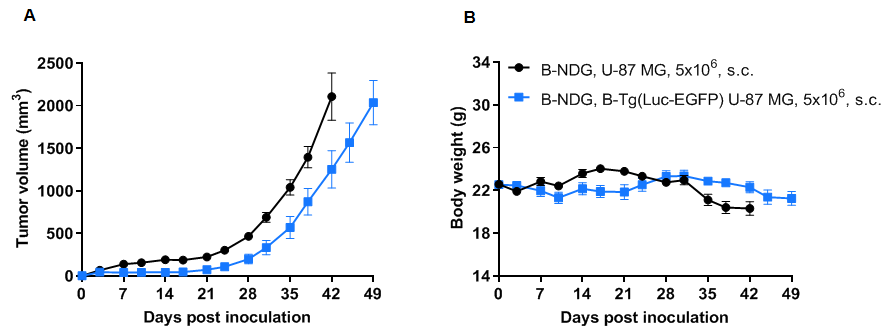
Subcutaneous tumor growth of B-Tg(Luc-EGFP) U-87 MG cells. B-Tg(Luc-EGFP) U-87 MG cells (5x10⁶) and wild-type U-87 MG cells (5x10⁶) were subcutaneously implanted into B-NDG mice (female, 7-9-week-old, n=6). Tumor volume and body weight were measured twice a week. (A) Average tumor volume. (B) Body weight. Volume was expressed in mm3 using the formula: V=0.5 X long diameter X short diameter2. Results indicate that B-Tg(Luc-EGFP) U-87 MG cells were able to establish tumors in vivo and can be used for efficacy studies. Values are expressed as mean ± SEM.
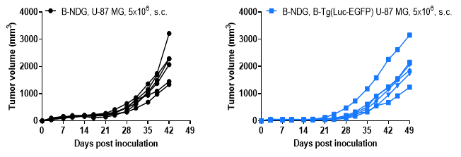
B-Tg(Luc-EGFP) U-87 MG tumor growth curves from individual mice. B-Tg(Luc-EGFP) U-87 MG cells (5x10⁶) and wild-type U-87 MG cells (5x10⁶) were subcutaneously implanted into B-NDG mice (female, 7-9-week-old, n=6). Results indicate that B-Tg(Luc-EGFP) U-87 MG cells were able to establish tumors in vivo and can be used for efficacy studies. Values are expressed as mean ± SEM.
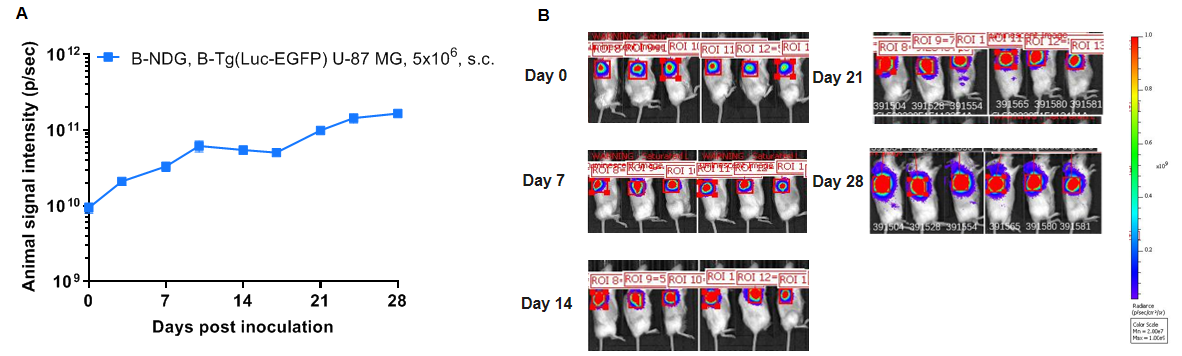
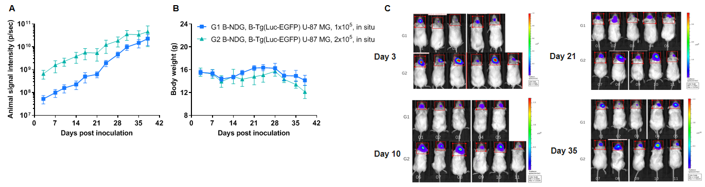
Growth kinetics of B-Tg(Luc-EGFP) U-87 MG tumors determined by bioluminescence imaging (BLI). B-Tg(Luc-EGFP) U-87 MG cells (1x10⁵, 2x10⁵) were injected into the brain of wild-type B-NDG mice (female, 8-week-old, G1 n=5, G2 n=6). Signal intensity and body weight were measured twice a week. (A) Signal intensity. (B) Body weight. (C) Raw bioluminescence images. These results indicate that B-Tg(Luc-EGFP) U-87 MG cells can be used for in vivo efficacy evaluation in brain orthotopic tumor models. Values are expressed as mean ± SEM.
