

• 321871
| Product name | B-Tg(Luc) RKO |
|---|---|
| Catalog number | 321871 |
| Tissue | Colon |
| Disease | Colon carcinoma |
| Species | Human |
| Application | B-Tg(Luc) RKO |
on this page
This B-Tg(Luc) RKO cell line expresses firefly luciferase as a marker of RKO cells. Fluorescence could be detected in B-Tg(Luc) RKO cells.
Gene targeting strategy for B-Tg(Luc) RKO cells. The exogenous promoter and luciferase coding sequence were inserted into the mouse genome randomly.
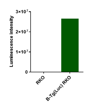
Luminescence signal intensity of B-Tg(Luc) RKO cells. Luminescence intensity was measured using the Bright-GloTM luciferase Assay (Promega, Cat E2610). B-Tg(Luc) RKO cells have a strong luminescence signal. The mix clone of B-Tg(Luc) RKO cells can be used for in vivo experiments.
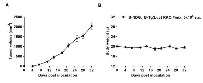
Subcutaneous homograft tumor growth of B-Tg(Luc) RKO cells. B-Tg(Luc) RKO cells (5x10⁶) were subcutaneously implanted into B-NDG mice (female, 7-week-old, n=5). Tumor volume and body weight were measured twice a week. (A) Average tumor volume ± SEM. (B) Body weight (Mean± SEM). Volume was expressed in mm3 using the formula: V=0.5 X long diameter X short diameter2. As shown in panel A, B-Tg(Luc) RKO cells were able to establish tumors in vivo and can be used for efficacy studies.
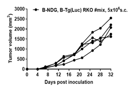
B-Tg(Luc) RKO tumor growth of individual mice. B-Tg(Luc) RKO cells (5x10⁶) were subcutaneously implanted into B-NDG mice (female, 7-week-old, n=5). As shown in panel, B-Tg(Luc) RKO cells were able to establish tumors in vivo and can be used for efficacy studies.
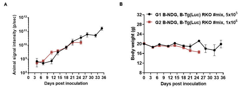
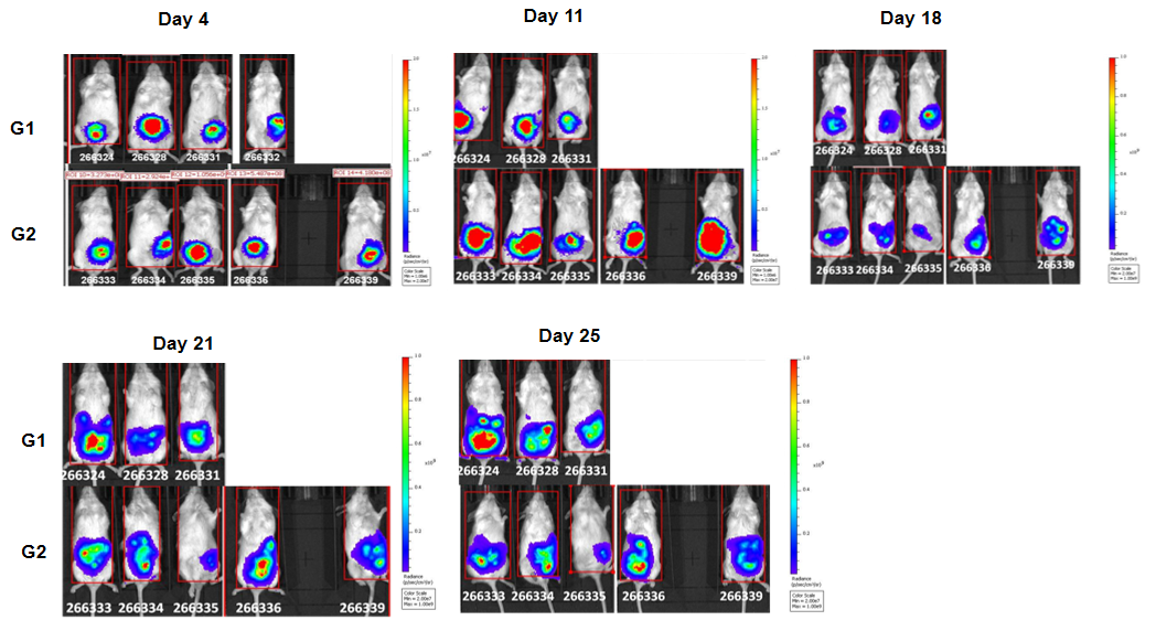
B-Tg(Luc) RKO cells (5x10⁵ ,1x106) were injected into the colon of wild-type B-NDG mice. Signal intensity and body weight were measured twice a week. (A) Imaging was performed twice a week. (B) Body weight (Mean ± SEM). B-Tg(Luc) RKO cells can be used for in vivo efficacy evaluation.
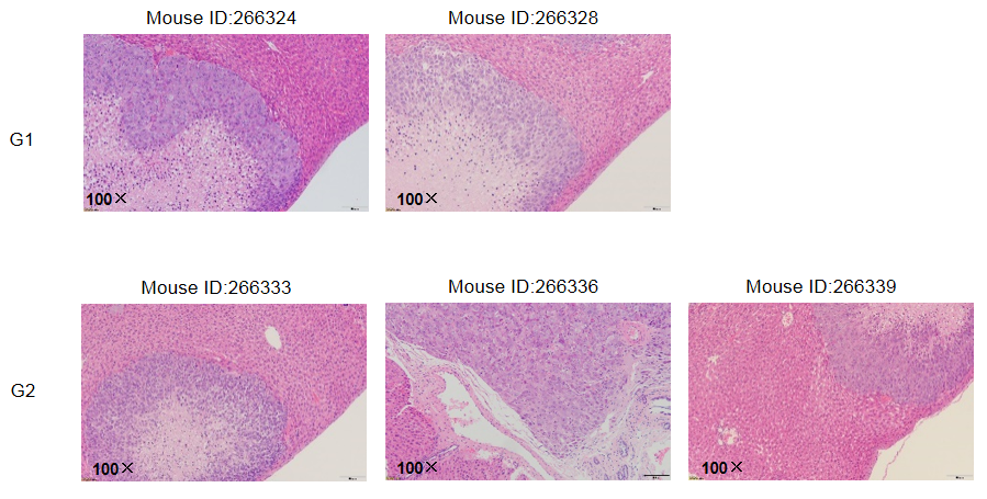
Tumor metastasis occurred in the liver of mice by HE.
