

C57BL/6-Faptm1(FAP)Bcgen/Bcgen • 110831
| Product name | B-hFAP mice |
|---|---|
| Catalog number | 110831 |
| Strain name | C57BL/6-Faptm1(FAP)Bcgen/Bcgen |
| Strain background | C57BL/6 |
| Aliases | FAPA; SIMP; DPPIV; FAPalpha |
Gene targeting strategy for B-hFAP mice. The exons 3-26 of mouse Fap gene that encode the extracellular domain were replaced by human FAP exons 3-26 in B-hFAP mice.
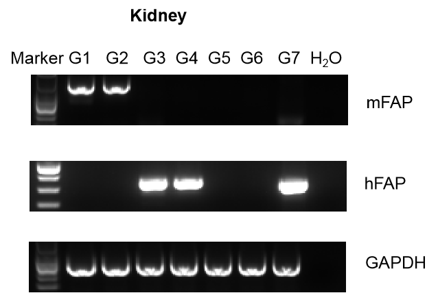
Strain specific analysis of FAP gene expression in wild-type C57BL/6 mice and B-hFAP mice by RT-PCR. Murine colon cancer MC38 cells (5×10⁵) and melanoma B16-F10 (2×10⁵) were specifically subcutaneously implanted into wild-type C57BL/6 mice, homozygous B-hFAP mice and homozygous B-Fap KO mice. B-CAG-hFAP MC38 cell line which over-expressing human FAP in MC38 cell line was used as a positive control. The mRNA was prepared from kidney tissue of the mice. Mouse Fap mRNA was detectable in kidney tissue of wild-type C57BL/6 mice. Human FAP mRNA was detectable in homozygous B-hFAP mice and B-CAG-hFAP MC38 cell line, but not in wild-type mice. Mouse Fap mRNA and human FAP mRNA were not detectable in homozygous B-Fap KO mice.
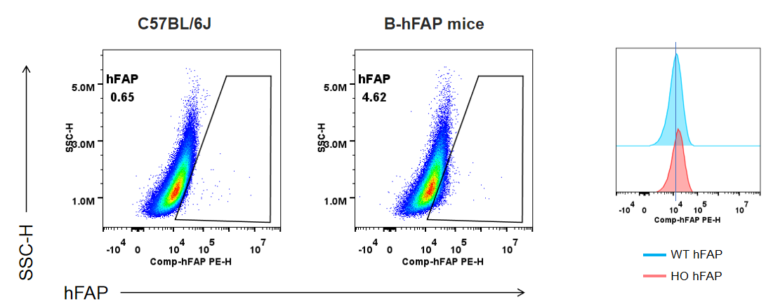
Strain specific FAP expression analysis in homozygous B-hFAP mice by flow cytometry. MEF cells was collected from wild-type C57BL/6JNifdc mice (+/+) and homozygous B-hFAP mice (H/H), and protein expression was analyzed with anti-human FAP antibody (R&D, FAB3715P-100) by flow cytometry. hFAP was detectable in MEF cells of homozygous B-hFAP mice.
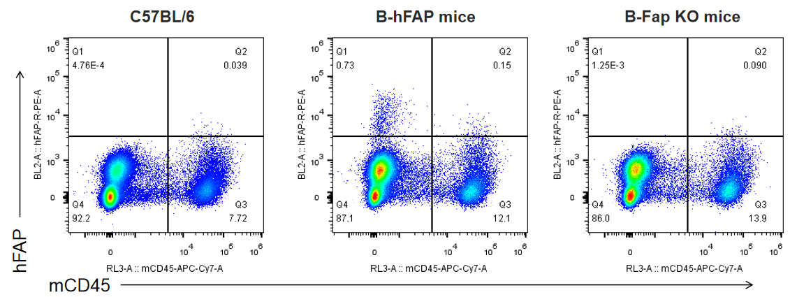
Strain specific analysis of FAP expression in wild-type C57BL/6 mice, homozygous B-hFAP mice and B-Fap KO mice by FACS. Murine melanoma B16-F10 (2×10⁵) were specifically subcutaneously implanted into wild-type C57BL/6 mice, homozygous B-hFAP mice and homozygous B-Fap KO mice. Tumor tissue was collected from the three mice, and analyzed by flow cytometry with species-specific anti-hFAP antibody (R&D, FAB3715P-100). Human FAP was exclusively detectable in CD45- cells of tumor tissue from homozygous B-hFAP mice but not in that cells from wild-type C57BL/6 mice or B-Fap KO mice.
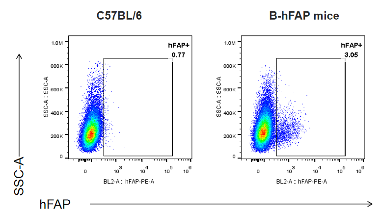
Strain specific analysis of FAP expression in wild-type C57BL/6 mice and homozygous B-hFAP mice by FACS. Murine Pan02 cells (2x10⁷) were specifically subcutaneously implanted into wild-type C57BL/6 mice and homozygous B-hFAP mice. Tumor tissue was collected from the three mice, and analyzed by flow cytometry with species-specific anti-hFAP antibody (R&D, FAB3715P-100). Human FAP was exclusively detectable in CD45- cells of tumor tissue from homozygous B-hFAP mice but not in that cells from wild-type C57BL/6 mice.
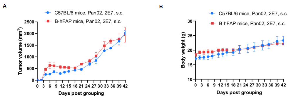
Subcutaneous homograft tumor growth of Pan02 cells. Pan02 cells (2x10⁷) were subcutaneously implanted into B-hFAP mice (female, n=3). Tumor volume and body weight were measured twice a week. (A) Average tumor volume ± SEM. (B) Body weight (Mean± SEM). Volume was expressed in mm3 using the formula: V=0.5 X long diameter X short diameter2. As shown in panel A, B-hFAP mice were able to establish tumors in vivo and can be used for efficacy studies.
