

NOD.CB17-Prkdcscid Il2rgtm1Bcgen B2mtm1Bcgen Fcgrttm1(B2m/Fcgrt)Bcgen /Bcgen • 110601
| Product name | B-NDG B2m KO mice plus |
|---|---|
| Catalog number | 110601 |
| Strain name | NOD.CB17-Prkdcscid Il2rgtm1Bcgen B2mtm1Bcgen Fcgrttm1(B2m/Fcgrt)Bcgen /Bcgen |
| Strain background | B-NDG |
| NCBI gene ID | 12010,14132 |
| Aliases | B2m: Ly-m11, beta2-m, beta2m; Fcgrt: FcRn |
| Application | Reduce PBMC reconstitution GVHD response and prolong experimental window |
Human PBMC-engrafted humanized mice are attractive models for in vivo analysis of human immune responses. We previously reported that human PBMCs could be engrafted in B-NDG mice successfully. However, because of severe xenogeneic graft versus host disease (xeno-GVHD) in these mice, there is a limited window for experiments. The onset of GvHD is directly related to the degree of engraftment of human T cells. The mismatches between human and mouse MHCs are the main cause of GvHD after human PBMC transplantation1. Data have shown that knockout of MHC class I and/or II molecules in mice attenuates GvHD and prolongs the window period for experiments after human PBMC transplantation.
MHC class I molecules consist of two subunits, α and β chains. The B2m (beta-2-microglobulin) gene encodes the β chain. Knockout of the B2m gene in mice can result in the inability to form intact MHC class I molecules and cannot be expressed on the cell surface to exert antigen presentation, thereby alleviating GvHD. However, B2M is also a subunit of FcRn, the Fc receptor for IgG. The structure of FcRn is similar to that of MHC class I molecules, consisting of the α chain encoded by the Fcgrt gene and the β chain encoded by the B2m gene. FcRn can bind to antibodies and prolong the half-life of antibodies. Loss of B2M expression results in a shortened half-life of the antibody in mice.
Biocytogen developed the B-NDG B2m KO plus mice, which knocked out the mouse B2m gene, while the mouse B2m and Fcrgt fusion gene were knocked in at the mouse Fcgrt gene locus. This mouse has a background of B-NDG mice and does not express MHC class I molecules on the cell surface. Compared with B-NDG mice, this mouse has no difference in the metabolism of IgG and is effective in delaying the development of GvHD. This model can be used to investigate the in vivo mechanism of xenogeneic graft versus host disease (GvHD) and to evaluate the efficacy of antitumor drugs using humanized mice reconstituted with human PBMCs.
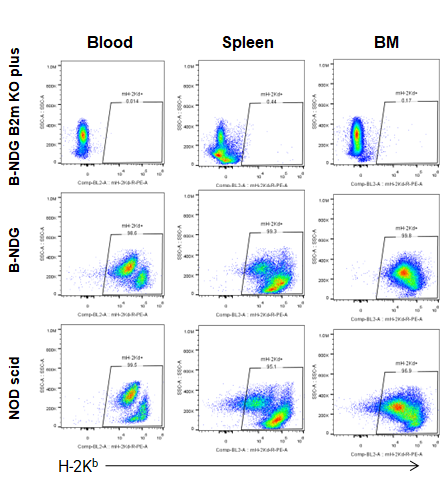
Flow cytometric analysis of MHC class l expression.
Blood cells, splenocytes, Bone marrow cells were collected from B-NDG B2m KO mice plus, B-NDG, NOD-scid mice (n=5) and analyzed by flow cytometry with MHC class I (H-2Kd). MHC class I were exclusively detectable in B-NDG, NOD-scid mice, but not in B-NDG B2m KO mice plus.
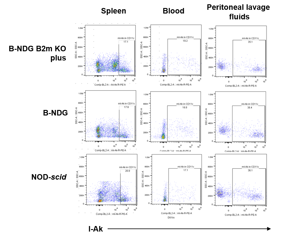
Flow cytometric analysis of MHC class lI expression in DC.
Blood cells, splenocytes, Peritoneal lavage fluids were collected from B-NDG B2m KO mice plus, B-NDG, NOD-scid mice (n=3) and analyzed by flow cytometry with MHC class II (mIA-IE). Single live cells were gated for DC (CD45+CD11b+CD11c+) population and used for further analysis as indicated here. MHC class II were detectable in DC subpopulations of B-NDG, NOD-scid mice and B-NDG B2m KO mice plus.
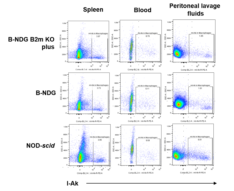
Flow cytometric analysis of MHC class lI expression in macrophages.
Blood cells, splenocytes, Peritoneal lavage fluids were collected from B-NDG B2m KO mice plus, B-NDG, NOD-scid mice (n=3) and analyzed by flow cytometry with MHC class II. Single live cells were gated for macrophages (CD45+CD11b+F4/80+) population and used for further analysis as indicated here. MHC class II were detectable in macrophage subpopulations of B-NDG, NOD-scid mice and B-NDG B2m KO mice plus.
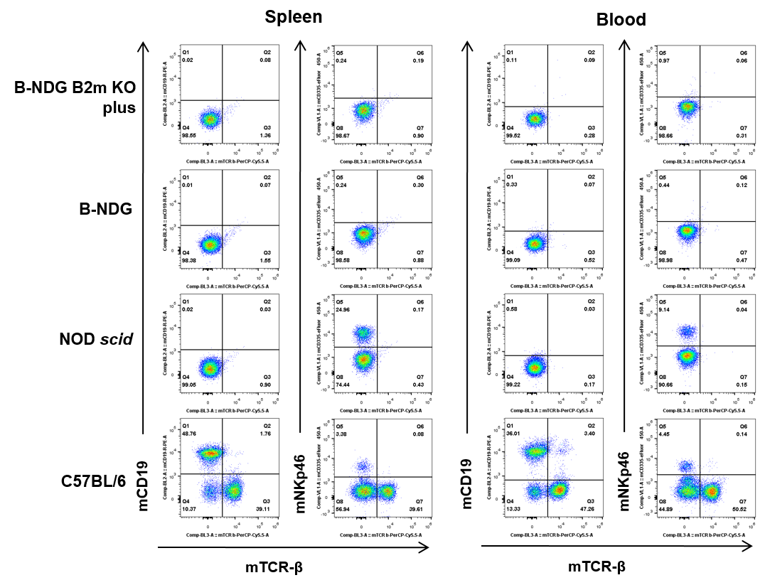
Flow cytometric analysis of T , B and NK cells.
Blood cells, splenocytes were collected from B-NDG B2m KO mice plus, B-NDG, NOD-scid and C57BL/6 mice (n=5) and analyzed by flow cytometry with T, B and NK cells. T, B and NK cells were not detectable in B-NDG B2m KO mice plus and B-NDG mice. T cells and B cells were undetectable and NK cells were detectable in NOD scid mice. T, B and NK cells were detectable in C57BL/6 mice.
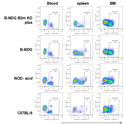
Flow cytometric analysis of Macrophages.
Blood cells, splenocyte, Bone marrow cells were collected from B-NDG B2m KO mice plus, B-NDG, NOD-scid and C57BL/6 mice (n=5) and analyzed by flow cytometry with Macrophages (mCD45+mCD11b+mF4-80+). Macrophages were detectable in B-NDG B2m KO mice plus, B-NDG, NOD-scid and C57BL/6 mice.
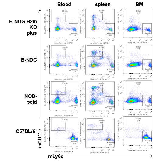
Flow cytometric analysis of DC cells and monocytes.
Blood cells, splenocytes, Bone marrow cells were collected from B-NDG B2m KO mice plus, B-NDG, NOD-scid and C57BL/6 mice (n=5) and analyzed by flow cytometry with DC cells (mCD45+mCD11b+mCD11c) and monocytes (mCD45+mCD11b+mLy6c). DC cells and monocytes were detectable in B-NDG B2m KO mice plus, B-NDG, NOD-scid and C57BL/6 mice.
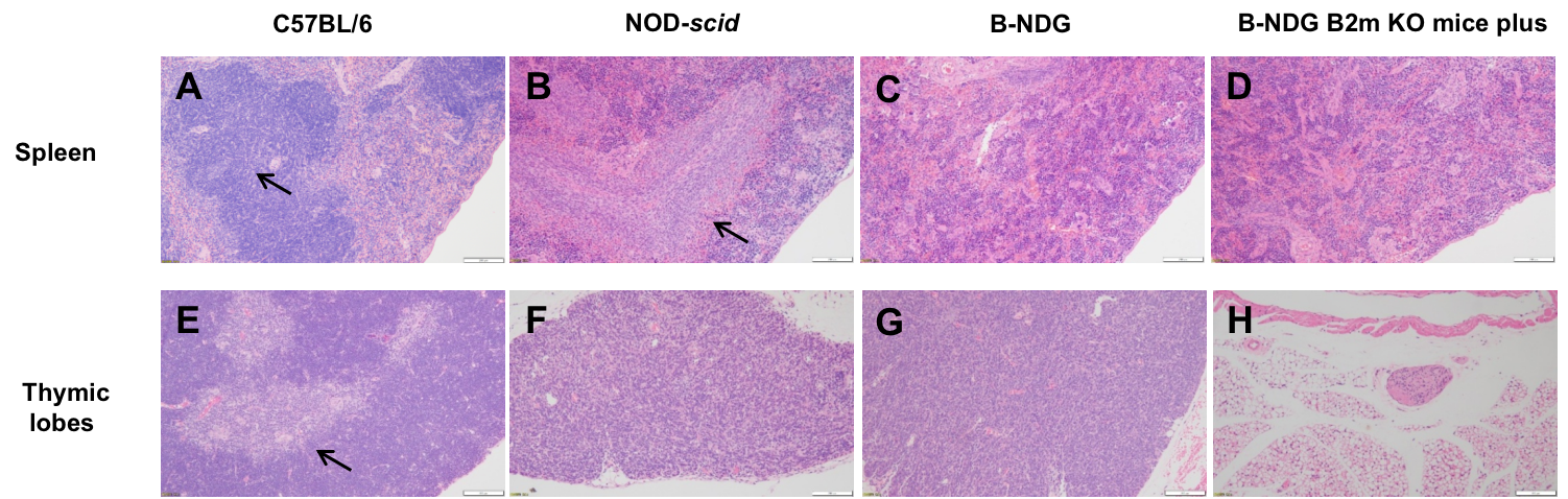
Histopathological sections of tissues from B-NDG B2m KO mice plus and control mice.
(A) Spleen from C57BL/6 has normal structure with well-defined follicles. (B) Spleen from NOD-scid show hypoplasia of white pulp. (C, D) Spleen from both B-NDG and B-NDG B2m KO mice plus show complete loss of follicular structure. (E) Thymic lobes from C57BL/6 have normal structure with a well-defined cortex. (F) Thymic lobes from NOD-scid mice are hypoplastic and lack a defined cortex.(g) Thymic lobes from B-NDG mice are severely hypoplastic and lack a defined cortex. (H) B-NDG B2m KO mice plus show no thymic lobes on the normal anatomy location.
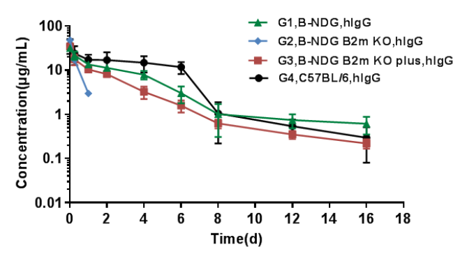
Pharmacokinetic characteristic of B-NDG B2m KO mice plus has no difference compared with wild-type mice.
Homozygous B-NDG, B-NDG B2m mice, B-NDG B2m KO mice plus and C57BL/6 mice were treated with human IgG (n=5). Blood samples were collected at different time point for the PK assay. The results showed that the PK results of B-NDG and B-NDG B2m KO mice plus groups were basically in line with the pharmacokinetic characteristics, with no difference compared with wild-type mice, and the drug concentration of B-NDG B2m KO mice group could not be measured at the time point 2 days later.
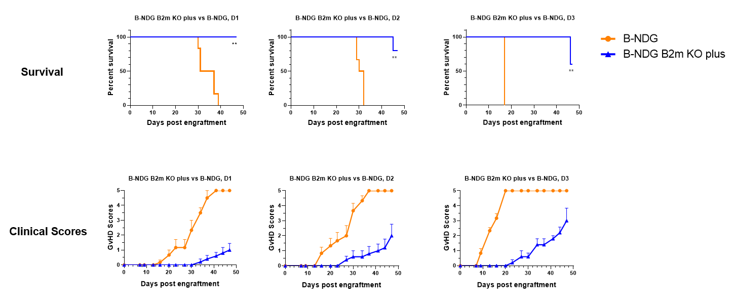
B-NDG B2m KO mice plus delay the onset and reduce the severity of GvHD compared to B-NDG mice.
B-NDG B2m KO mice plus (11-week-old female, n=5) and B-NDG (10-week-old female, n=6) mice were engrafted i.v. with 2E6 human PBMC from three healthy donors (D1-3) on day 0. Clinical signs of GvHD were scored approximately twice a week. Values were expressed as mean ± SEM. **: P<0.01. The result showed that B-NDG B2m KO mice plus significantly extend survival compared with B-NDG mice in the human PBMC induced GvHD model.
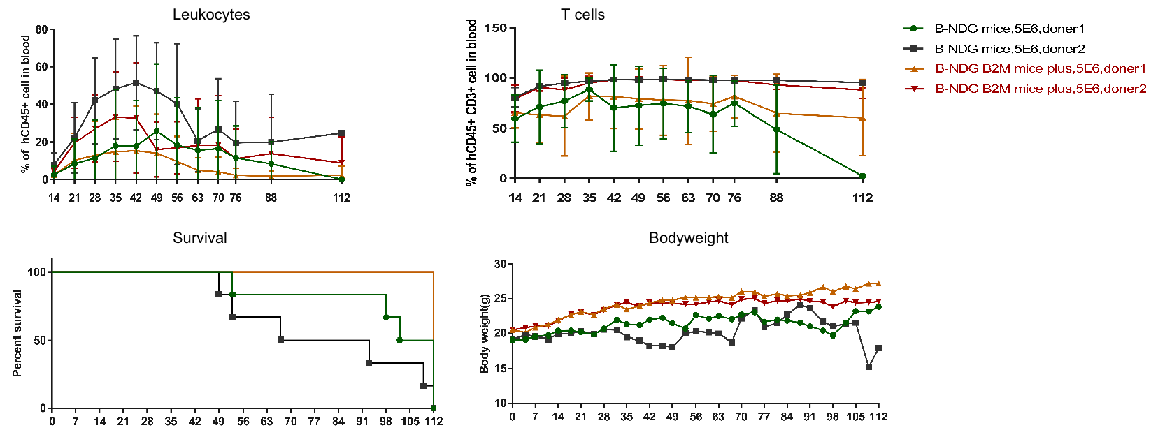
B-NDG B2m KO mice plus are well suited for human immune system engraftment.
Human PBMC (5E6) were intravenous implanted into homozygote B-NDG B2m KO mice plus and B-NDG mice (female, 7-week-old, n=6). Representative flow cytometric analysis of PBMCs from mice after engraftment with human PBMC. B-NDG B2m KO mice plus show a little change in body weight and exhibit longer survival compared with B-NDG. The result showed that human PBMC engrafted humanized mice model was successfully constructed.

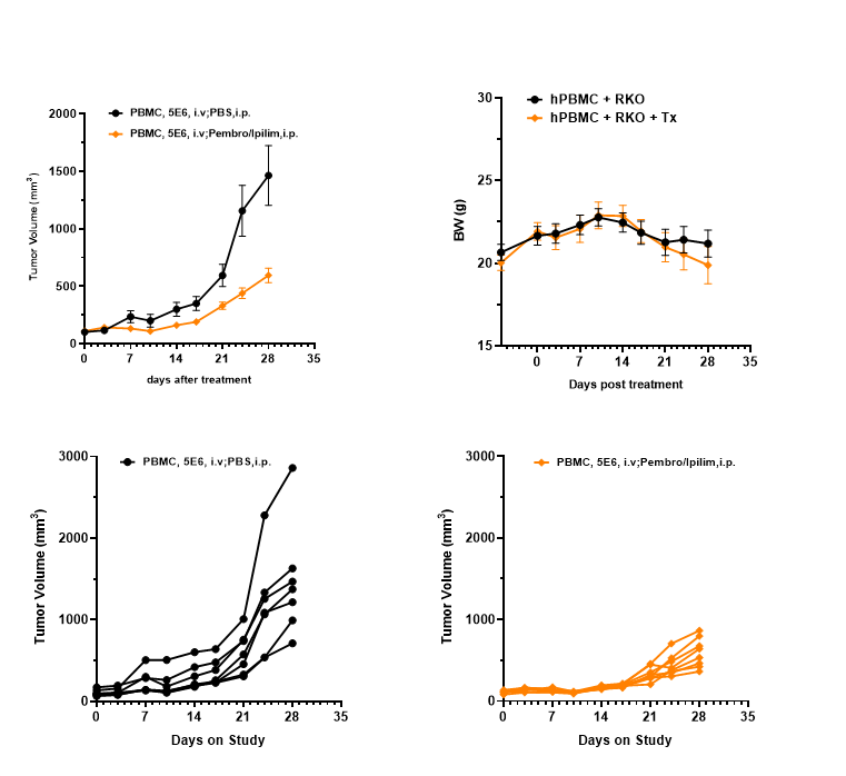
Antitumor activity of the combination of Pembrolizumab (anti PD-1) and Ipilimumab (anti CTLA-4) in B-NDG B2m KO mice plus.
(A) Schematic diagram of PBMC RKO model and the anti-tumor assay, Human RKO cells (5E6) were subcutaneously implanted after human PBMCs (10E6) injected into B-NDG B2m mice plus (female, 9 week-old, n=7/8). The animals were grouped into control and treatment and tumor size were measured every two weeks. (B) Tumor volume and body weight changes during treatment. The combination of Pembrolizumab and Ipilimumab shows significant tumor inhibitory effects. The results indicate that B-NDG B2m mice plus reconstituted with PBMCs provides a powerful preclinical model for in vivo evaluation of antibodies. Values are expressed as mean ± SEM.
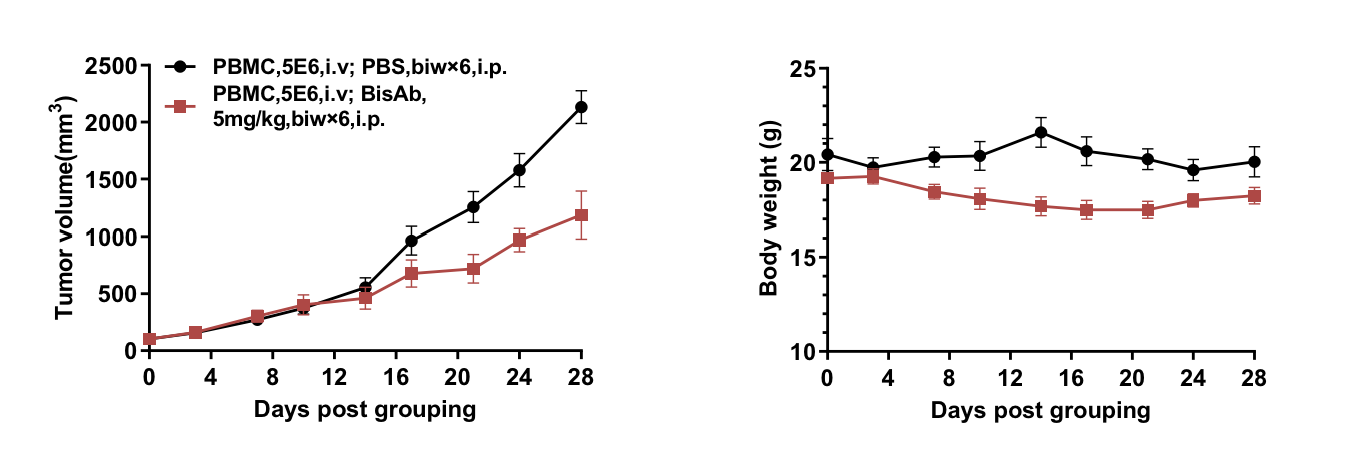
B-NDG B2m mice plus reconstituted with PBMCs were used for anti PD-1×PD-L1 bispecific antibody efficacy studies
Human RKO cells (5E6) were subcutaneously implanted after human PBMCs (5E6) implanted into B-NDG B2m mice plus (female, 9 week-old, n=6). The animals were grouped into control and treatment when the tumor size was approximately 100 mm3. (A) Anti human PD-1×PD-L1 bispecific antibody inhibited RKO tumor growth in human PBMC reconstituted B-NDG B2m mice plus. (B) Body weight changes during treatment. Anti PD-1×PD-L1bispecific antibody shows significant tumor inhibitory effects. The results indicate that B-NDG B2m mice plus reconstituted with PBMCs provides a powerful preclinical model for in vivo evaluation of antibodies. Values are expressed as mean ± SEM.
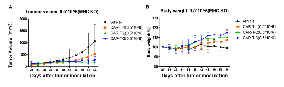
Antitumor activity of CAR-T therapy in B-NDG B2m KO mice plus.
Human NCI-H226 (A) (1E7) were implanted into B-NDG B2m KO mice plus. (A) Mice were grouped when tumor size was approximately 150±50 mm3. at which time they were treated with CAR-T cells (5E5) with schedules indicated in panel. (B) Body weight changes during treatment. The results showed that CAR-T cells differently inhibited tumor growth in B-NDG B2m KO mice plus. B2m KO mice plus is a powerful model for human CAR-T cells efficacy study. Values are expressed as mean ± SEM.
