

NOD.CB17-Prkdcscid Il2rgtm1Bcgen B2mtm2(B2M/HLA-A2.1)Bcgen/Bcgen • 110599
| Product name | B-NDG HLA-A2.1 mice |
|---|---|
| Catalog number | 110599 |
| Strain name | NOD.CB17-Prkdcscid Il2rgtm1Bcgen B2mtm2(B2M/HLA-A2.1)Bcgen/Bcgen |
| Strain background | B-NDG |
| Aliases | B2m: Ly-m11, beta2-m, beta2m; HLA-A: HLAA |
| Application | Vaccine study |
The B2m gene (Exon1 to Exon3) of mouse was replaced by the sequence encompassing the human B2M CDS and HLA-A2.1 gene.
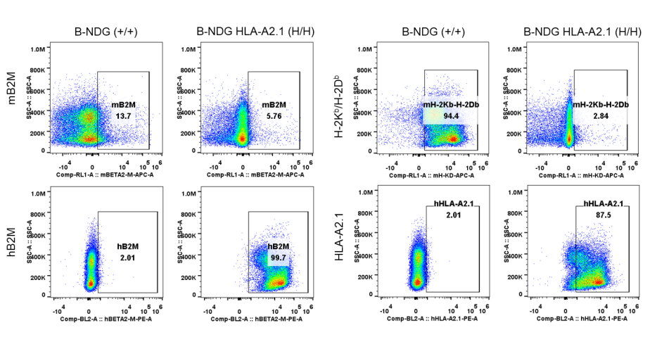
HLA-A2.1 expression analysis in homozygous B-NDG HLA-A2.1 mice by flow cytometry. Splenocytes were collected from B-NDG mice (+/+) and homozygous B-NDG HLA-A2.1 mice (H/H), and analyzed by flow cytometry with strain specific antibodies. Mouse B2M and H-2Kb/H-2Db were detectable in B-NDG mice but not in B-NDG HLA-A2.1 mice. Human B2M and HLA-A2.1 was exclusively detectable in homozygous B-NDG HLA-A2.1 mice but not in B-NDG mice.
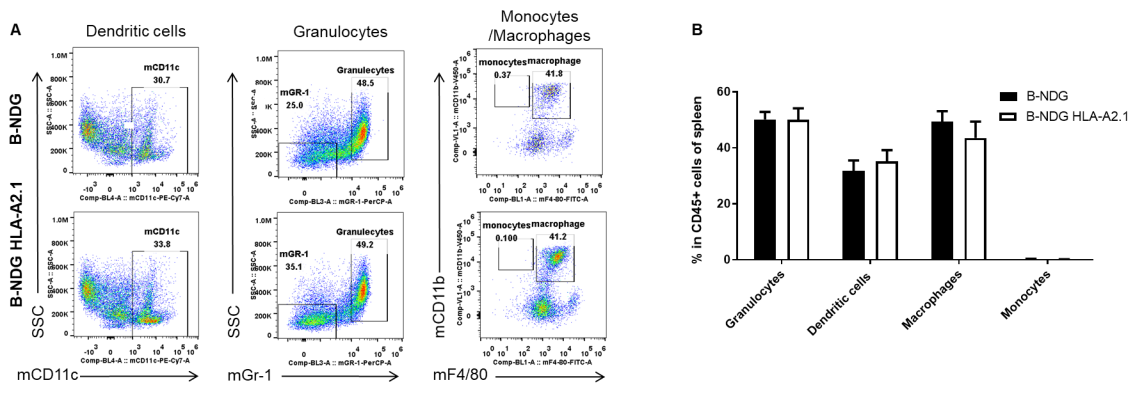
Analysis of spleen leukocyte subpopulations by FACS. Splenocytes were isolated from female B-NDG mice and B-NDG HLA-A2.1 mice (n=3, 6-week-old). Flow cytometry analysis of the splenocytes was performed to assess leukocyte subpopulations. A. Representative FACS plots. Single live cells were gated for the CD45+ population and used for further analysis as indicated here. B. Results of FACS analysis. Percentages of dendritic cells, granulocytes, monocytes and macrophages in homozygous B-NDG HLA-A2.1 mice were similar to those in the B-NDG mice, demonstrating that B2M and HLA-A2.1 humanized do not change the overall development, differentiation or distribution of these cell types in spleen. Values are expressed as mean ± SEM.
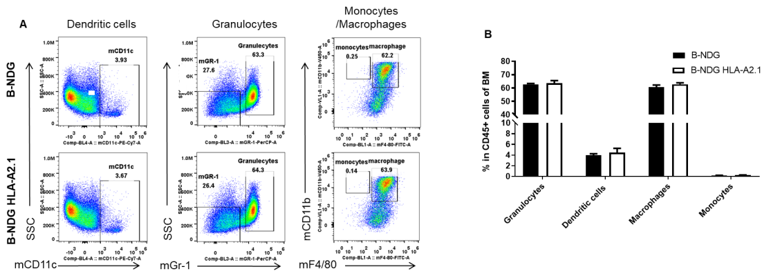
Analysis of bone marrow leukocyte subpopulations by FACS. Bone marrow was isolated from female B-NDG mice and B-NDG HLA-A2.1 mice (n=3, 6-week-old). Flow cytometry analysis was performed to assess leukocyte subpopulations. A. Representative FACS plots. Single live cells were gated for the CD45+ population and used for further analysis as indicated here. B. Results of FACS analysis. Percentages of dendritic cells, granulocytes, monocytes and macrophages in homozygous B-NDG HLA-A2.1 mice were similar to those in the B-NDG mice, demonstrating that B2M and HLA-A2.1 humanized do not change the overall development, differentiation or distribution of these cell types in bone marrow. Values are expressed as mean ± SEM.
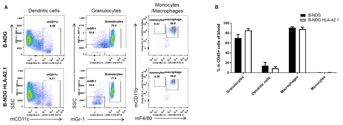
Analysis of blood leukocyte subpopulations by FACS. Blood was isolated from female B-NDG mice and B-NDG HLA-A2.1 mice (n=3, 6-week-old). Flow cytometry analysis was performed to assess leukocyte subpopulations. A. Representative FACS plots. Single live cells were gated for the CD45+ population and used for further analysis as indicated here. B. Results of FACS analysis. Percentages of dendritic cells, granulocytes, monocytes and macrophages in homozygous B-NDG HLA-A2.1 mice were similar to those in the B-NDG mice, demonstrating that B2M and HLA-A2.1 humanized do not change the overall development, differentiation or distribution of these cell types in blood. Values are expressed as mean ± SEM.
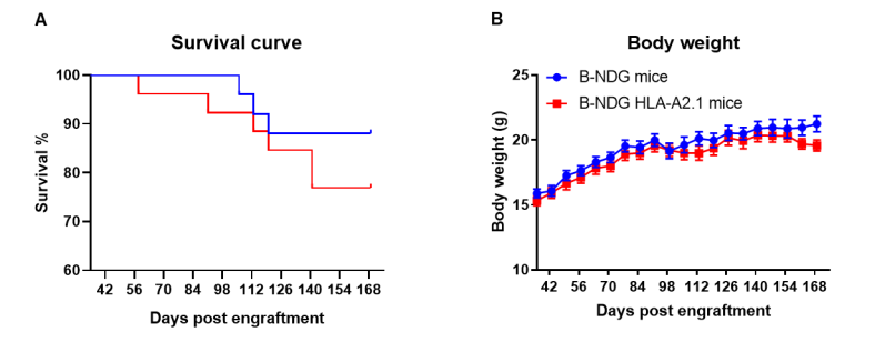
Human CD34+ HSCs were successfully engrafted in B-NDG HLA-A2.1 mice. Human CD34+ HSCs were respectively engrafted in new born mice of B-NDG HLA-A2.1 mice and B-NDG mice after radiated with 0.8 Gy. (A) B-NDG HLA-A2.1 mice showed slightly lower survival rate than B-NDG mice, but no statistically significant difference. (B) Body weight.
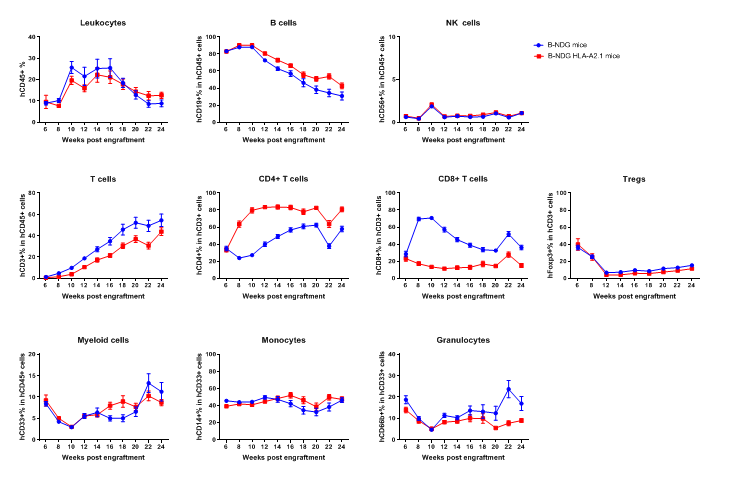
Human CD34+ HSCs were successfully engrafted in B-NDG HLA-A2.1 mice. Human CD34+ HSCs were respectively engrafted in new born mice of B-NDG HLA-A2.1 mice and B-NDG mice after radiated with 0.8 Gy. Human leukocytes were analyzed by flow cytometry. Results showed that although the proportion of hCD8 + T cells in B-NDG HLA-A2.1 mice was significantly lower than that in B-NDG mice, it remains sustained at around 18% within 24 weeks of reconstitution. The proportion of hCD4 + T cells in B-NDG HLA-A2.1 mice was significantly higher than that in B-NDG mice. Proportions of other reconstituted cell types in B-NDG HLA-A2.1 mice were similar to that in B-NDG mice.
