

NOD.CB17-Prkdcscid Il2rgtm1Bcgen Sirpatm1(SIRPA)Bcgen/Bcgen • 110604
| Product name | B-NDG hSIRPA mice |
|---|---|
| Catalog number | 110604 |
| Strain name | NOD.CB17-Prkdcscid Il2rgtm1Bcgen Sirpatm1(SIRPA)Bcgen/Bcgen |
| Strain background | B-NDG |
| NCBI gene ID | 19261 (Human) |
| Aliases | SIRPα (signal regulatory protein alpha), BIT, CD172A, MFR, MYD-1, P84, PTPNS1, SHPS1, SIRP |
| Application | Maintain progenitor capacity |
Signal regulatory protein α (SIRPα) is a transmembrane protein with an extracellular region comprising three Ig-like domains and a cytoplasmic region containing immunoreceptor tyrosine-based inhibition motifs which mediate binding of the protein tyrosine phosphatases SHP1 and SHP2. SIRPα is especially abundant in myeloid cells such as macrophages and dendritic cells(DC), whereas it is expressed at very low levels in T, B ,NK, and NK T cells. SIRPα inhibits phagocytosis in macrophages upon interacting with its ligand CD47, which is commonly upregulated on the surface of malignant cells. Thus, antibodies that block the CD47-SIRPα interaction should enhance macrophage phagocytosis in the tumor microenvironment and inhibit tumor growth, making anti-SIRPα antibodies promising tools for cancer immunotherapy.
Biocytogen developed the B-NDG hSIRPA mice, and the targeting strategy was that the exon 2 of mouse Sirpα gene that encode the extracellular domain were replaced by human SIRPα exon 2 in B-NDG hSIRPA mice. This mouse combines a B-NDG mouse background (completely lacking mature T, B and NK cells and were deficient in cytokine signaling) and expresses human SIRPα protein extracellular domain. In homozygous mice, mouse SIRPα were absent and only human protein expression was detected. B-NDG hSIRPA mice paired with genetically modified Raji-luc cancer cells were used to evaluate the efficacy of antibodies targeting SIRPα. Anti-human SIRPα antibodies were efficacious in controlling tumor growth in B-NDG hSIRPA mice. Humanized B-NDG hSIRPA mice are a promising in vivo efficacy model for the development of SIRPα antibodies that can be advanced to human clinical trials.
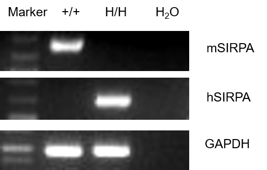
Strain specific analysis of SIRPA gene expression in B-NDG mice and B-NDG hSIRPA (H/H) mice by RT-PCR. Mouse Sirpa mRNA was detectable only in splenocytes of B-NDG mice (+/+). Human SIRPA mRNA was detectable only in homozygous B-NDG hSIRPA mice (H/H), but not in B-NDG mice.
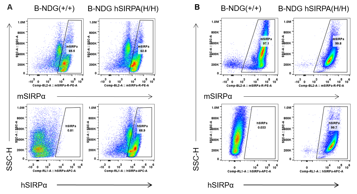
Strain specific SIRPα expression analysis in homologous B-NDG hSIRPA mice by flow cytometry. Splenocytes (A) and peritoneal lymphocyte (B) from B-NDG and homozygous B-NDG hSIRPA (H/H) mice were analyzed by flow cytometry with anti-SIRPα antibodies. Mouse SIRPα was detectable in B-NDG and homozygous B-NDG hSIRPA mice. This anti-mouse SIRPα antibody also cross reacts with human SIRPα. Human SIRPα were exclusively detectable in homozygous B-NDG hSIRPA but not B-NDG mice.
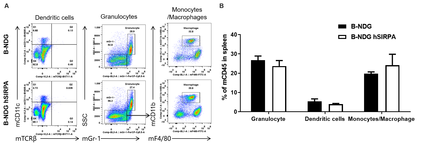
Analysis of spleen leukocyte subpopulations by FACS. Splenocytes were isolated from male B-NDG and B-NDG hSIRPA mice (n=3, 7-week-old). Flow cytometry analysis of the splenocytes was performed to assess leukocyte subpopulations. A. Representative FACS plots. Single live cells were gated for CD45 population and used for further analysis as indicated here. B. Results of FACS analysis. Percent of Monocyte, DC and macrophage cells in homozygous B-NDG hSIRPA mice were similar to those in the B-NDG mice, demonstrating that introduction of hSIRPα in place of its mouse counterpart does not change the overall development, differentiation or distribution of these cell types in spleen.
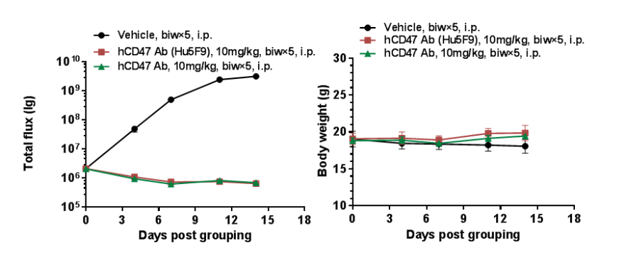
Antitumor activity of anti-human CD47 antibodies in B-NDG hSIRPA mice. (A) Human B-luciferase-GFP Raji cells (B lymphocytes) (5.0E+05) were inoculated into homozygous B-NDG hSIRPA mice (female, 5-week-old, n=5). Mice were grouped when the fluorescence intensity reached 1.0E+06 , at which time they were treated with anti-human CD47 antibody with doses and schedules indicated in panel. (B) Body weight changes during treatment. As shown in panel A, anti-human CD47 antibody was efficacious in controlling tumor growth in this model. Values are expressed as mean ± SEM.
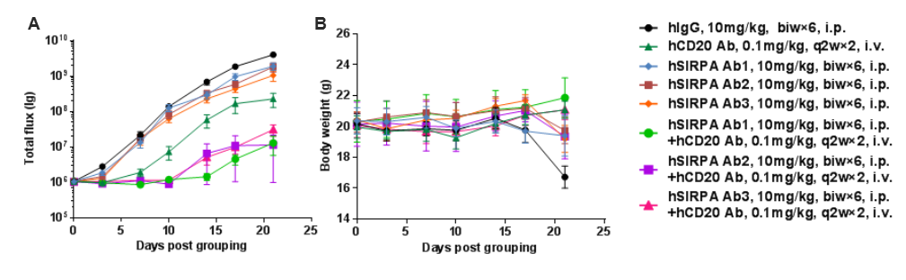
Antitumor activity of anti-human SIRPA antibody combined with anti-human CD20 antibody in B-NDG hSIRPA mice. (A) hSIRPA antibody combined with hCD20 antibody inhibited B-luciferase-GFP Raji tumor growth in B-NDG hSIRPA mice. Human B-luciferase-GFP Raji cells (B lymphocytes) (5.0E+05) were inoculated into homozygous B-NDG hSIRPA mice (female, 8-week-old, n=6). Mice were grouped when the fluorescence intensity reached approximately 1E6 p/sec, at which time they were treated with hSIRPA and hCD20 antibodies with doses and schedules indicated in panel A. (B) Body weight changes during treatment. As shown in panel A, combination of hSIRPA and hCD20 antibodies shows more inhibitory effects than individual groups. Values are expressed as mean ± SEM.
