
on this page
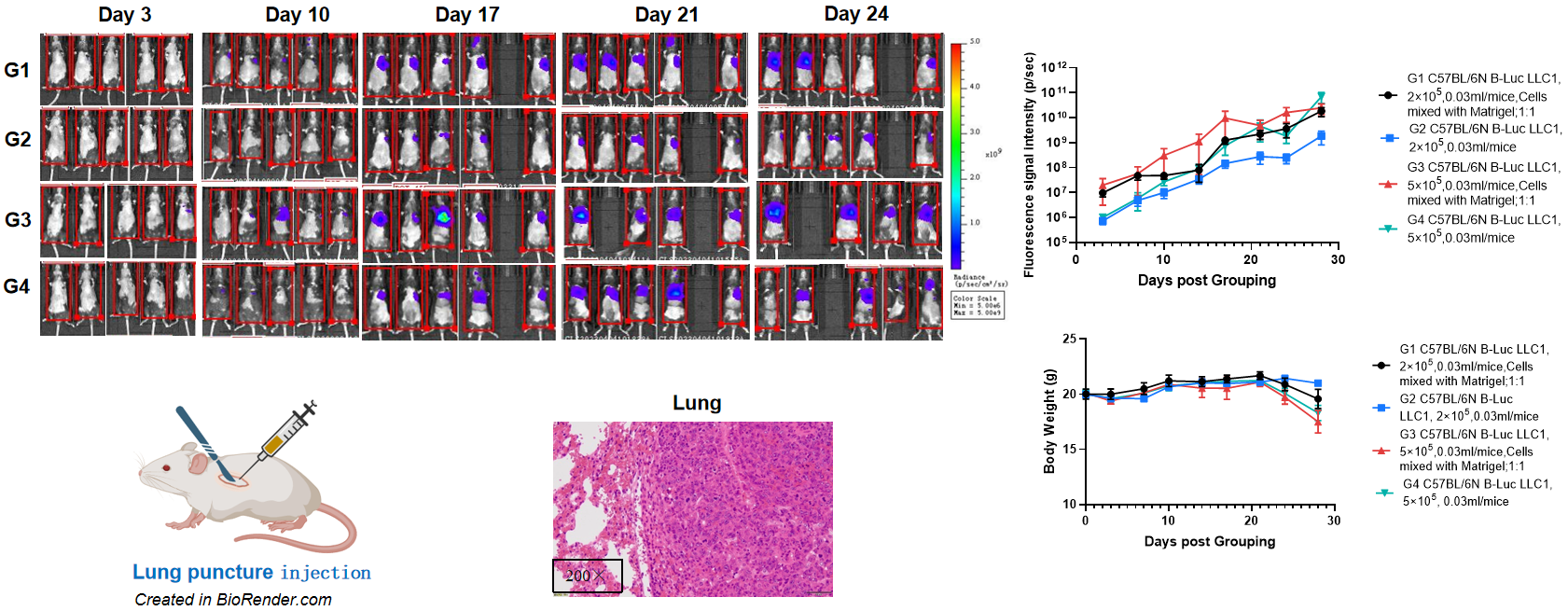
Establishment of orthotopic lung cancer model.
The C57BL/6 mice(F, n=5) were inoculated with a volume of 200 μL of B-Luc LLC1 cell of two different doses (2×10⁵, 5×10⁵) with or without matrigel by lung puncture injection , and the body weight and tumor fluorescence signal of the mice were recorded weekly. The results showed that the fluorescence signal gradually increased and body weight of mice started decrease from day21. This indicated that the cell line was successfully constructed as an orthotopic tumor model. H&E staining showed that there was obvious tumor formation in the lung.
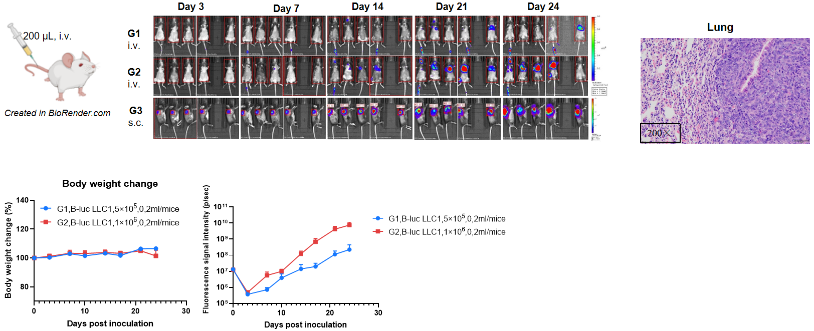
Establishment of orthotopic lung cancer model.
The C57BL/6 mice were inoculated by tail vein with a volume of 200 μL of B-Luc LLC1 cell suspension (5×10⁵, 1×10⁶), and the body weight and tumor fluorescence signal of the mice were recorded weekly. The results showed that the fluorescence signal gradually increased and body weight of mice did not change over time. Right panel showed H&E staining of liver tumors.. This indicated that the cell line was successfully constructed as an orthotopic tumor model.
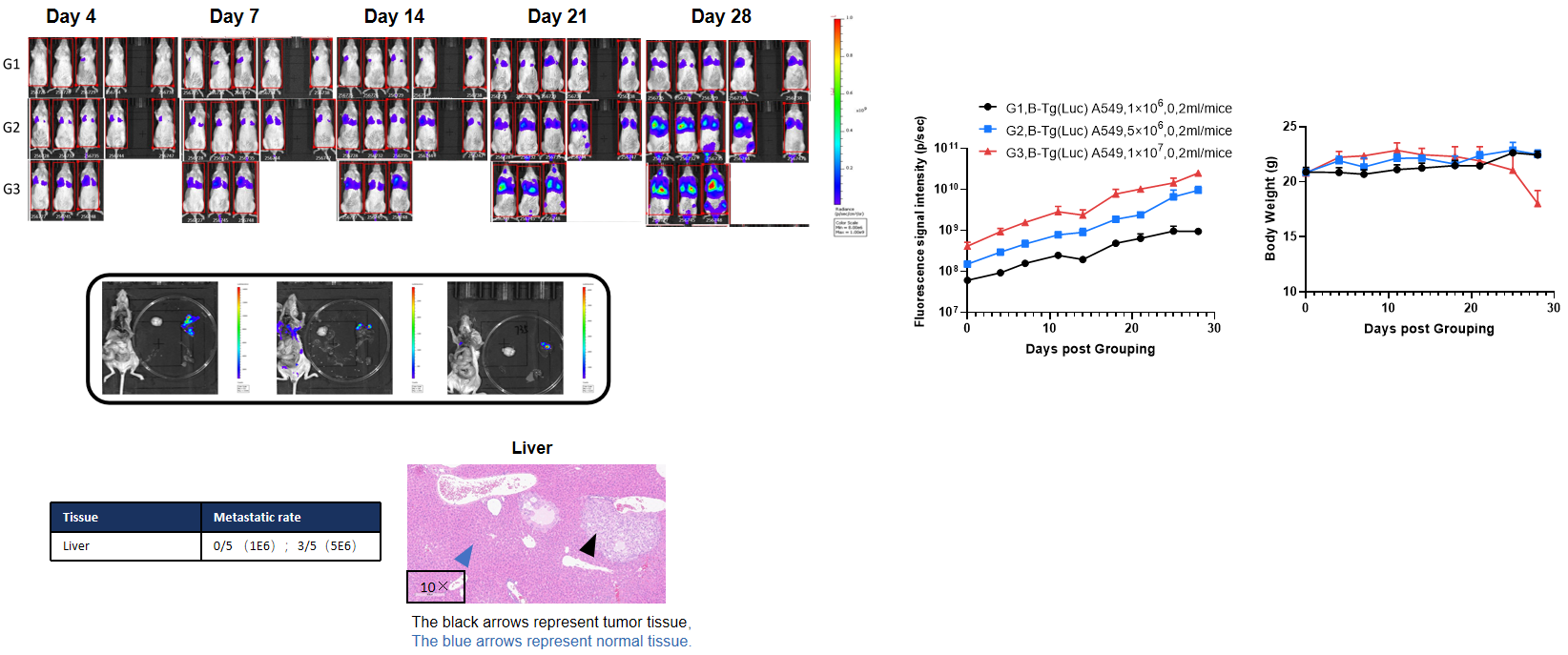
Establishment of orthotopic lung cancer model.
The different densities of B-Tg(Luc) A549 cell suspension (1×10⁶, 5×10⁶, 1×10⁷) was injected into B-NDG mice by tail vein injection, and the live imaging were observed with a small-animal live imaging instrument, The tumors keep steady growing and the body weight of mice did not change significantly during the experiment, but the G3 group showed a significant decrease in body weight due to high-seeding density. Mice were dissected at the end of the experiment, it can be seen that obvious fluorescent signals were observed in the tissues where the tumors were removed, implying that metastasis of the tumors occurred, and the pathological results showed that tumors metastasized in the liver.
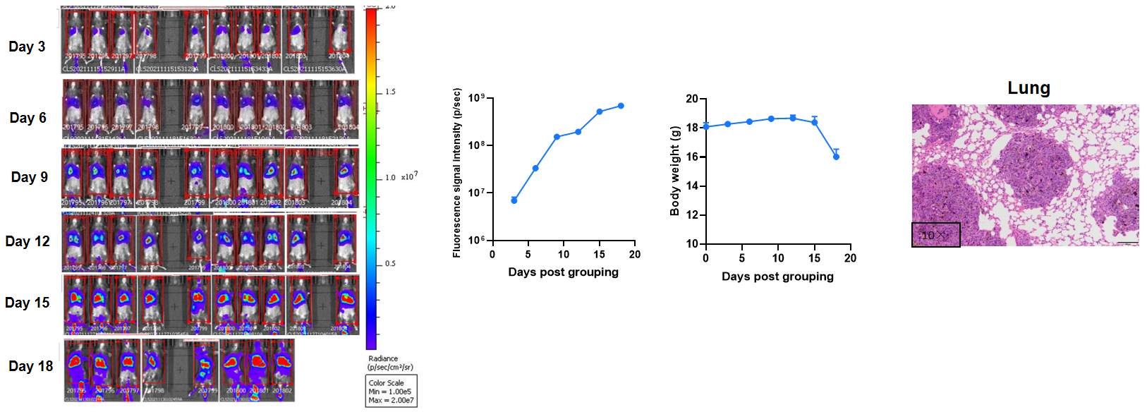
Establishment of B-luc B16-F10 tumor metastasis model.
The C57BL/6 mice (F, n=10) were inoculated by tail vein with a volume of 200 μL of B-luc B16-F10 cell suspension (2×10⁵), and the body weight and tumor fluorescence signal of the mice were recorded weekly. The results showed that the tumors keep steady growing and the body weight decreased from day15 during the experiment. H&E staining showed that there was obvious tumor formation in the lung. These results indicated that the B-luc B16-F10 cell line was successfully constructed as lung metastasis model.
