

C57BL/6-Camk2atm1(icre,EGFP)Bcgen/Bcgen • 110780
| Product name | B-Camk2a-iCre-EGFPmice |
|---|---|
| Catalog number | 110780 |
| Strain name | C57BL/6-Camk2atm1(icre,EGFP)Bcgen/Bcgen |
| Strain background | C57BL/6 |
| NCBI gene ID | 12322 (Mouse) |
| Official symbol | Camk2a, calcium/calmodulin-dependent protein kinase II alpha |
| Chromosome | 18 |
| Application | Function researchof genes;Function research of synaptic plasticity and Long TermPotentiation (LTP) in hippocampal networks |
Mice homozygous for the Camk2a-iCre are viable, fertile, normal in size and do not display any gross physical or behavioral abnormalities. In this strain, iCre recombinase expression is under the control of Camk2a promoter. When crossed with a strain containing a loxP-site flanked sequence of interest, Cre-mediated recombination will result in deletion of the floxed sequence in the offspring.
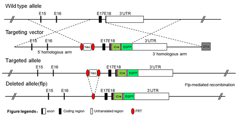
A targeting vector was designed to insert an P2A-iCre-P2A-EGFP cassette before the calcium/calmodulin-dependent protein kinase II alpha (Camk2a) stop codon. A Frt-flanked neomycin resistance (Neo) gene was inserted into the intron16-17 of Camk2a gene. The targeting vector was electroporated into C57BL/6-derived embryonic stem (ES) cells. Correctly targeted ES cells were injected into blastocysts and resulting chimeric mice were bred to FLP transgene mice to delete the neomycin selection cassette. FLP transgene was removed by crossing with wild-type C57BL/6 mice. These mice were maintained on a C57BL/6 background.
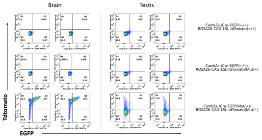
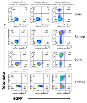
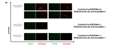
The flow results showed that the mouse brain could express ICRE and function normally. The single positive cells of TDTomato were found in testis, heart, liver, spleen, lung and kidney by flow cytometry.The experiment results with Jackson Lab B6. Cg - Tg (Camk2a - cre) STL T29-1 / J (Stock No: 005359 | T29-1) multiple organ test in mice to express the result of the agreement.The IF results of the fluorescent microscopic imaging of the mouse brain showed that the red fluorescent protein of TDTomato was successfully expressed in the mouse brain, and the expression was significantly increased in some areas, especially in the structure of the dentate gyrus, which was highly expressed.
