 Protein expression analysis
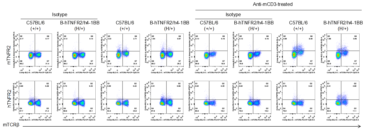
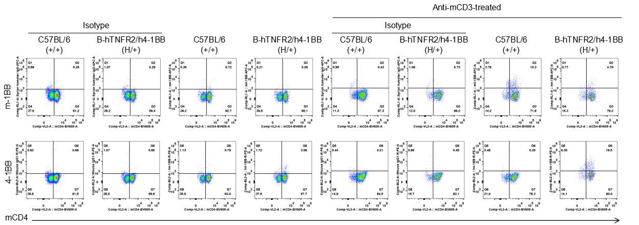
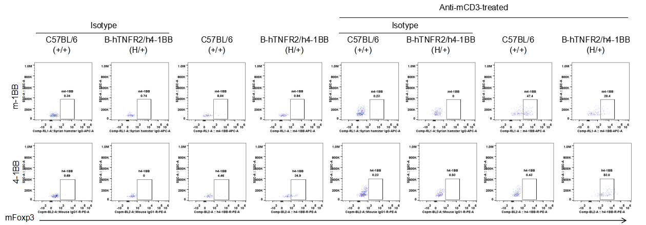
Strain specific TNFR2 expression analysis in heterozygous B-h4-1BB/hTNFR2 mice plus by flow cytometry. Splenocytes were collected from wild-type mice (+/+) and heterozygous B-h4-1BB/hTNFR2 mice plus (H/+) stimulated with anti-CD3ε in vivo (7.5 μg/mice, 24 hours, i.p.), and analyzed by flow cytometry with species-specific anti-TNFR2 antibody. Mouse TNFR2 was detectable in wild-type mice and heterozygous B-h4-1BB/hTNFR2 mice plus, but human TNFR2 was only detectable in heterozygous B-h4-1BB/hTNFR2 mice plus.
Strain specific TNFR2 expression analysis in heterozygous B-h4-1BB/hTNFR2 mice plus by flow cytometry. Splenocytes were collected from wild-type mice (+/+) and heterozygous B-h4-1BB/hTNFR2 mice plus (H/+) stimulated with anti-CD3ε in vivo (7.5 μg/mice, 24 hours, i.p.), and analyzed by flow cytometry with species-specific anti-TNFR2 antibody. Mouse TNFR2 was detectable in wild-type mice and heterozygous B-h4-1BB/hTNFR2 mice plus, but human TNFR2 was only detectable in heterozygous B-h4-1BB/hTNFR2 mice plus.
Strain specific TNFR2 expression analysis in heterozygous B-h4-1BB/hTNFR2 mice plus by flow cytometry. Splenocytes were collected from wild-type mice (+/+) and heterozygous B-h4-1BB/hTNFR2 mice plus (H/+) stimulated with anti-CD3ε in vivo (7.5 μg/mice, 24 hours, i.p.), and analyzed by flow cytometry with species-specific anti-TNFR2 antibody. Mouse TNFR2 was detectable in wild-type mice and heterozygous B-h4-1BB/hTNFR2 mice plus, but human TNFR2 was only detectable in heterozygous B-h4-1BB/hTNFR2 mice plus.
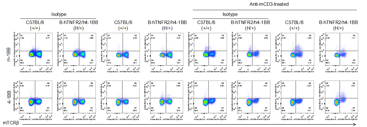
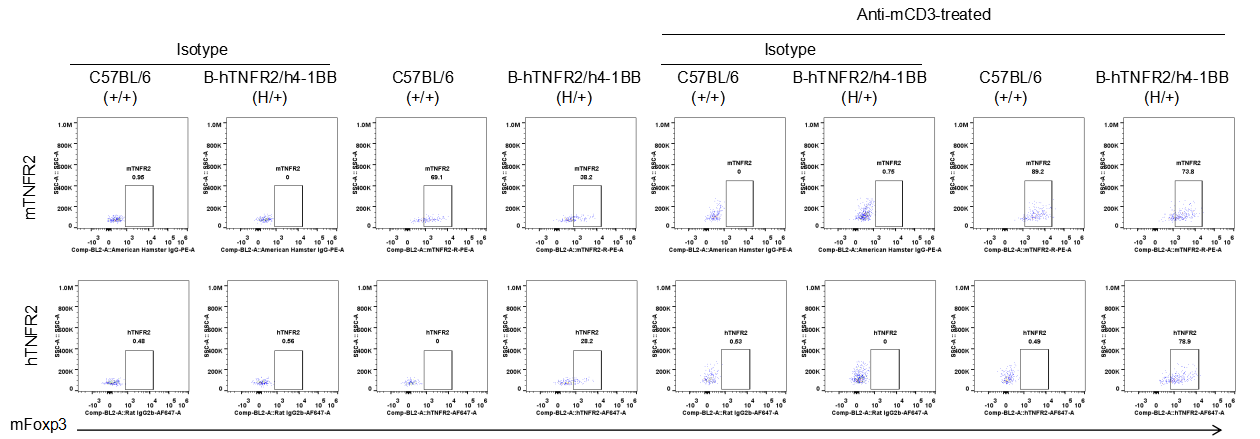
Strain specific 4-1BB expression analysis in heterozygous B-h4-1BB/hTNFR2 mice plus by flow cytometry. Splenocytes were collected from wild-type mice (+/+) and heterozygous B-h4-1BB/hTNFR2 mice plus (H/+) stimulated with anti-CD3ε in vivo (7.5 μg/mice, 24 hours, i.p.), and analyzed by flow cytometry with species-specific anti-4-1BB antibody. Mouse 4-1BB was detectable in wild-type mice and heterozygous B-h4-1BB/hTNFR2 mice plus, but human 4-1BB was only detectable in heterozygous B-h4-1BB/hTNFR2 mice plus.
Strain specific 4-1BB expression analysis in heterozygous B-h4-1BB/hTNFR2 mice plus by flow cytometry. Splenocytes were collected from wild-type mice (+/+) and heterozygous B-h4-1BB/hTNFR2 mice plus (H/+) stimulated with anti-CD3ε in vivo (7.5 μg/mice, 24 hours, i.p.), and analyzed by flow cytometry with species-specific anti-4-1BB antibody. Mouse 4-1BB was detectable in wild-type mice and heterozygous B-h4-1BB/hTNFR2 mice plus, but human 4-1BB was only detectable in heterozygous B-h4-1BB/hTNFR2 mice plus.
Strain specific 4-1BB expression analysis in heterozygous B-h4-1BB/hTNFR2 mice plus by flow cytometry. Splenocytes were collected from wild-type mice (+/+) and heterozygous B-h4-1BB/hTNFR2 mice plus (H/+) stimulated with anti-CD3ε in vivo (7.5 μg/mice, 24 hours, i.p.), and analyzed by flow cytometry with species-specific anti-4-1BB antibody. Mouse 4-1BB was detectable in wild-type mice and heterozygous B-h4-1BB/hTNFR2 mice plus, but human 4-1BB was only detectable in heterozygous B-h4-1BB/hTNFR2 mice plus.