

BALB/cCrSIcNifdc-H2-Ea1tm1(HLA-DRA1*1.1-I-Ea)Bcgen H2-Eb1tm1(HLA-DRB1*4.1-I-Eb)Bcgen H2-Atm1Bcgen H2-Eb2tm1Bcgen/Bcgen • 112720
| Product name | B-hHLA-DRB1*4.1 mice(C) |
|---|---|
| Catalog number | 112720 |
| Strain name | BALB/cCrSIcNifdc-H2-Ea1tm1(HLA-DRA1*1.1-I-Ea)Bcgen H2-Eb1tm1(HLA-DRB1*4.1-I-Eb)Bcgen H2-Atm1Bcgen H2-Eb2tm1Bcgen/Bcgen |
| Strain background | BALB/cCrSlcNifdc |
| NCBI gene ID | 3122, 3123 |
| Aliases | HLA-DRA1; SS1, DRB1, HLA-DRB, HLA-DR1B |
Gene targeting strategy for B-hHLA-DRB1*4.1 mice(C). The exons 2-4 of mouse I-Ea1 gene that encode the extracellular and transmembrane domain were replaced by human counterparts in B-hHLA-DRB1*4.1 mice(C). The exons 2-4 of mouse I-Eb1 gene that encode the extracellular and transmembrane domain were replaced by human counterparts in B-hHLA-DRB1*4.1 mice(C). The mouse I-Aa, I-Ab and I-Eb2 were knocked out in B-hHLA-DRB1*4.1 mice(C).
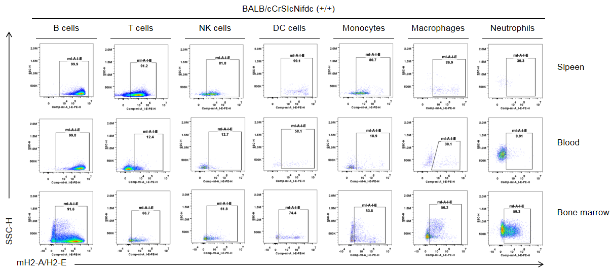
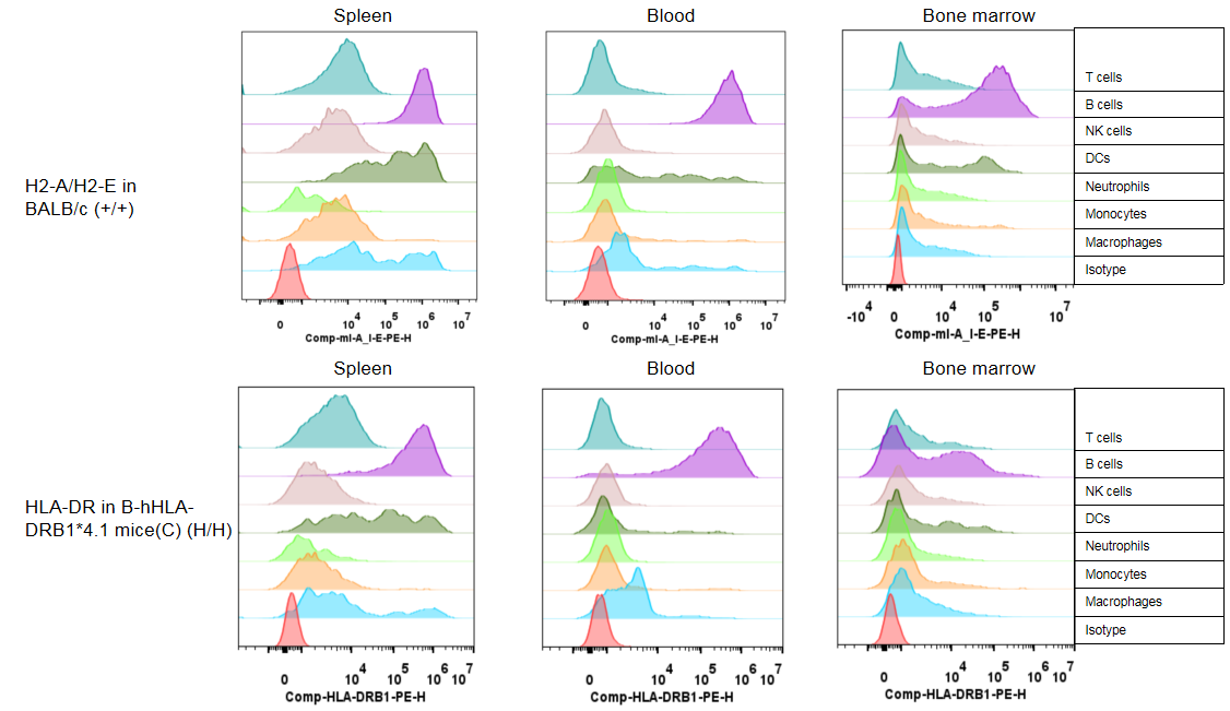
Strain specific mH2-A/H2-E expression analysis in wild-type BALB/cCrSIcNifdc mice by flow cytometry. Splenocytes, blood cells and bone marrow cells were collected from wild-type BALB/cCrSIcNifdc mice (+/+), and analyzed by flow cytometry with species-specific anti-mouse H2-A/H2-E antibody(Biolegend, 107607). Mouse H2-A/H2-E was only detectable in immune cells of wild-type mice but not in immune cells of B-hHLA-DRB1*4.1 mice(C) (The page 6).
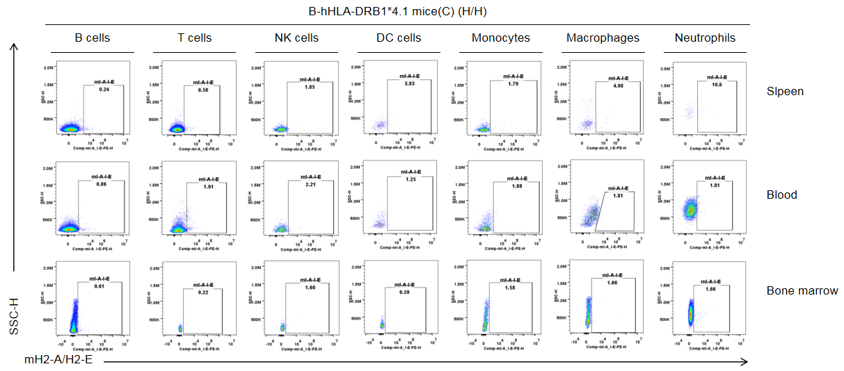
Strain specific mH2-A/H2-E expression analysis in B-hHLA-DRB1*4.1 mice(C) by flow cytometry. Splenocytes, blood cells and bone marrow cells were collected from B-hHLA-DRB1*4.1 mice(C), and analyzed by flow cytometry with species-specific anti-mouse H2-A/H2-E antibody(Biolegend, 107607). Mouse H2-A/H2-E was not detectable in immune cells of B-hHLA-DRB1*4.1 mice(C).
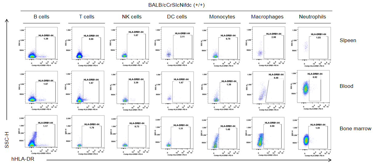
Strain specific HLA-DR expression analysis in wild-type BALB/cCrSIcNifdc mice by flow cytometry. Splenocytes, blood cells and bone marrow cells were collected from wild-type BALB/cCrSIcNifdc mice (+/+), and analyzed by flow cytometry with species-specific anti-HLA-DRB1 antibody(Biolegend, 307624). Human HLA-DR was not detectable in immune cells of wild-type mice but exclusively in immune cells of B-hHLA-DRB1*4.1 mice(C) (The page 8).
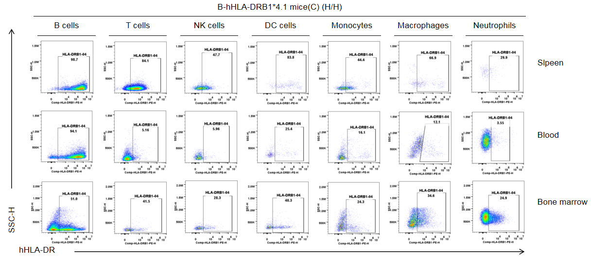
Strain specific HLA-DR expression analysis in B-hHLA-DRB1*4.1 mice(C) by flow cytometry. Splenocytes, blood cells and bone marrow cells were collected from B-hHLA-DRB1*4.1 mice(C), and analyzed by flow cytometry with species-specific anti-HLA-DR antibody(Biolegend, 307624). Human HLA-DR was only detectable in immune cells of B-hHLA-DRB1*4.1 mice(C).
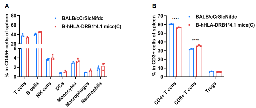
Frequency of leukocyte subpopulations in the spleen by flow cytometry. Splenocytes were isolated from wild-type BALB/cCrSlcNifdc mice and homozygous B-hHLA-DRB1*4.1 mice(C) (Female,8-week-old, n=3). A. Flow cytometry analysis of the splenocytes was performed to assess the frequency of leukocyte subpopulations. B. Frequencies of T cell subpopulations. Frequencies of NK cells, DCs, neutrophils, monocytes, macrophages, and Tregs in B-hHLA-DRB1*4.1 mice(C) were similar to those in BALB/cCrSlcNifdc mice. The frequency of B cells was significantly increased, while the frequency of T cells was significantly decreased. The frequency of CD8+ T cells in B-hHLA-DRB1*4.1 mice(C) was significantly increased, while the frequency of CD4+ T cells were significantly decreased, demonstrating that humanized mice modification affects the proportion of T cell subtypes in the spleen. Values are expressed as mean ± SEM. Significance was determined by two-way ANOVA test. *P < 0.05, **P < 0.01, ***P < 0.001.
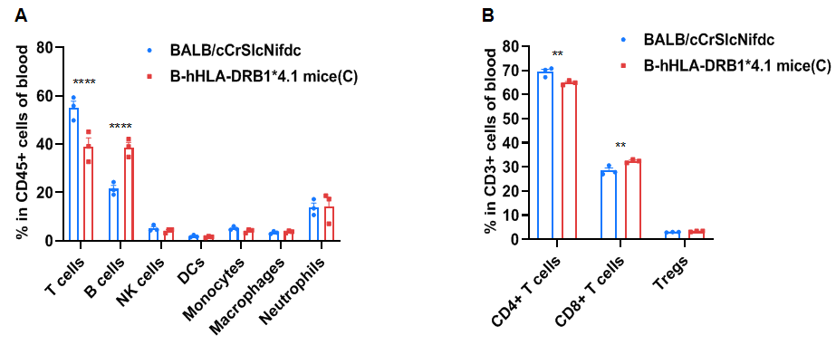
Frequency of leukocyte subpopulations in the blood by flow cytometry. Blood cells were isolated from wild-type BALB/cCrSlcNifdc mice and homozygous B-hHLA-DRB1*4.1 mice(C) (Female,8-week-old, n=3). A. Flow cytometry analysis of the blood was performed to assess the frequency of leukocyte subpopulations. B. Frequency of T cell subpopulations. Frequencies of NK cells, DCs, neutrophils, monocytes, macrophages, and Tregs in B-hHLA-DRB1*4.1 mice(C) were similar to those in BALB/cCrSlcNifdc mice. The frequency of B cells was significantly increased, while the frequency of T cells was significantly decreased. The frequency of CD8+ T cells in B-hHLA-DRB1*4.1 mice(C) was significantly increased, while the frequency of CD4+ T cells was significantly decreased, demonstrating that humanized mice modification affects the proportion of T cell subtypes in the blood. Values are expressed as mean ± SEM. Significance was determined by two-way ANOVA test. *P < 0.05, **P < 0.01, ***P < 0.001.
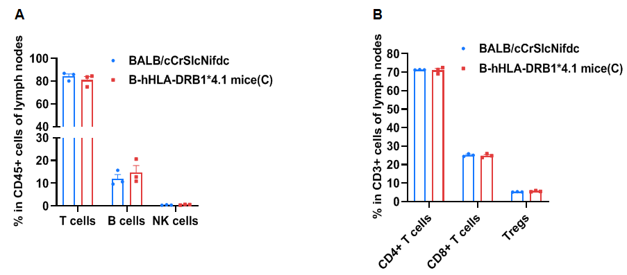
Frequency of leukocyte subpopulations in the lymph node by flow cytometry. The Lymph node cells were isolated from wild-type BALB/cCrSlcNifdc mice and homozygous B-hHLA-DRB1*4.1 mice(C) (Female,8-week-old, n=3). A. Flow cytometry analysis of the lymph node was performed to assess the frequency of leukocyte subpopulations. B. Frequency of T cell subpopulations. Frequencies of T cells, B cells, NK cells, CD4+ T cells, CD8+ T cells, and Tregs in B-hHLA-DRB1*4.1 mice(C) were similar to those in BALB/cCrSlcNifdc mice, demonstrating that humanization of HLA-DRB1*4.1 does not change the frequency or distribution of these cell types in the lymph node. Values are expressed as mean ± SEM. Significance was determined by two-way ANOVA test. *P < 0.05, **P < 0.01, ***P < 0.001.
