
C57BL/6-Lair1tm1(LAIR1)Bcgen/Bcgen • 111660
| Product name | B-hLAIR1 mice |
|---|---|
| Catalog number | 111660 |
| Strain name | C57BL/6-Lair1tm1(LAIR1)Bcgen/Bcgen |
| Strain background | C57BL/6N |
| Aliases | LAIR1 (CD305; LAIR-1) |
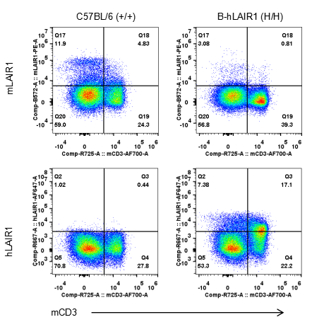
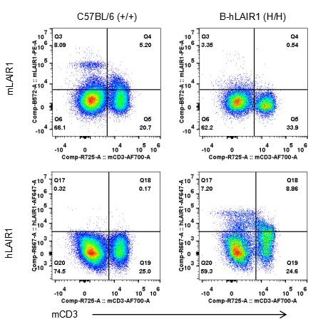
Strain specific LAIR1 expression analysis in homozygous B-hLAIR1 mice by flow cytometry. T cells from spleen were collected from wild-type C57BL/6 mice (+/+) and homozygous B-hLAIR1 mice (H/H), and analyzed by flow cytometry with species-specific anti-LAIR1 antibody. Mouse LAIR1 was detectable in wild-type mice. Human LAIR1 was exclusively detectable in homozygous B-hLAIR1 mice but not in wild-type mice.
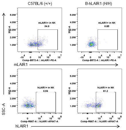
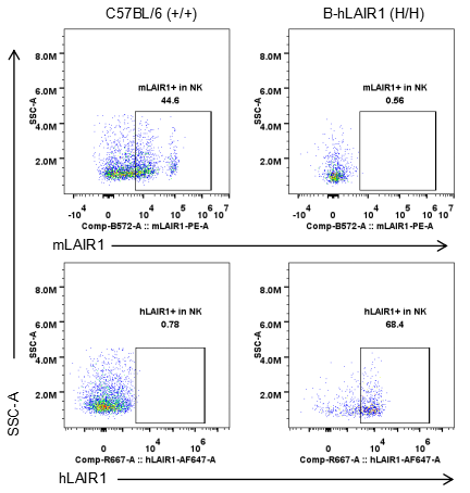
Strain specific LAIR1 expression analysis in homozygous B-hLAIR1 mice by flow cytometry. NK cells from spleen were collected from wild-type C57BL/6 mice (+/+) and homozygous B-hLAIR1 mice (H/H), and analyzed by flow cytometry with species-specific anti-LAIR1 antibody. Mouse LAIR1 was detectable in wild-type mice. Human LAIR1 was exclusively detectable in homozygous B-hLAIR1 mice but not in wild-type mice.
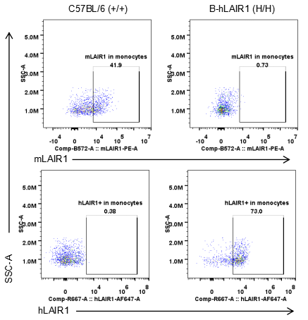
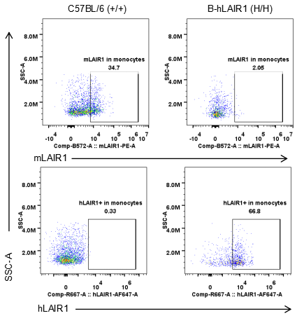
Strain specific LAIR1 expression analysis in homozygous B-hLAIR1 mice by flow cytometry. Monocytes from spleen were collected from wild-type C57BL/6 mice (+/+) and homozygous B-hLAIR1 mice (H/H), and analyzed by flow cytometry with species-specific anti-LAIR1 antibody. Mouse LAIR1 was detectable in wild-type mice. Human LAIR1 was exclusively detectable in homozygous B-hLAIR1 mice but not in wild-type mice.
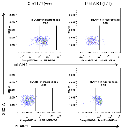
Strain specific LAIR1 expression analysis in homozygous B-hLAIR1 mice by flow cytometry. Macrophages from spleen were collected from wild-type C57BL/6 mice (+/+) and homozygous B-hLAIR1 mice (H/H), and analyzed by flow cytometry with species-specific anti-LAIR1 antibody. Mouse LAIR1 was detectable in wild-type mice. Human LAIR1 was exclusively detectable in homozygous B-hLAIR1 mice but not in wild-type mice.
Strain specific LAIR1 expression analysis in homozygous B-hLAIR1 mice by flow cytometry. T cells from blood were collected from wild-type C57BL/6 mice (+/+) and homozygous B-hLAIR1 mice (H/H), and analyzed by flow cytometry with species-specific anti-LAIR1 antibody. Mouse LAIR1 was detectable in wild-type mice. Human LAIR1 was exclusively detectable in homozygous B-hLAIR1 mice but not in wild-type mice.
Strain specific LAIR1 expression analysis in homozygous B-hLAIR1 mice by flow cytometry. NK cells from blood were collected from wild-type C57BL/6 mice (+/+) and homozygous B-hLAIR1 mice (H/H), and analyzed by flow cytometry with species-specific anti-LAIR1 antibody. Mouse LAIR1 was detectable in wild-type mice. Human LAIR1 was exclusively detectable in homozygous B-hLAIR1 mice but not in wild-type mice.
Strain specific LAIR1 expression analysis in homozygous B-hLAIR1 mice by flow cytometry. Monocytes from blood were collected from wild-type C57BL/6 mice (+/+) and homozygous B-hLAIR1 mice (H/H), and analyzed by flow cytometry with species-specific anti-LAIR1 antibody. Mouse LAIR1 was detectable in wild-type mice. Human LAIR1 was exclusively detectable in homozygous B-hLAIR1 mice but not in wild-type mice.
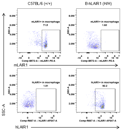
Strain specific LAIR1 expression analysis in homozygous B-hLAIR1 mice by flow cytometry. Macrophages from blood were collected from wild-type C57BL/6 mice (+/+) and homozygous B-hLAIR1 mice (H/H), and analyzed by flow cytometry with species-specific anti-LAIR1 antibody. Mouse LAIR1 was detectable in wild-type mice. Human LAIR1 was exclusively detectable in homozygous B-hLAIR1 mice but not in wild-type mice.
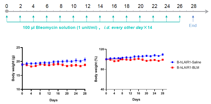
Mouse body weight changes over the study period. Body weight and percentage changes of mice during the experiment in B-hLAIR1 mice. N=6 mice per group.
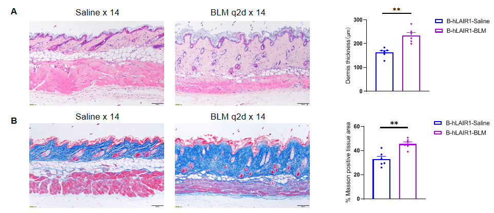
Dermis thickness of skin. Representive image of HE staining and dermis thickness change in B-hLAIR1 mice. N=6 mice per group.Positive area of Masson staining. Representive image of Masson staining and percentage change of Masson positive tissue area in B-hLAIR1 mice. N=6 mice per group
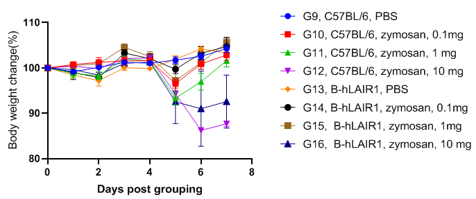
Body weight changes in Zymosan induced peritonitis models. The body weight percentage relative to the day of grouping (Day 0) are illustrated. Two way ANOVA with multiple comparison test, all groups compared with each other. Values are expressed as mean ± SEM. Significances are separately listed in the table (only meaningful comparisons are listed, i.e. significant comparison between groups with single independent variable in design).
Body weights for animals in G1-G8 were not illustrated as modeling and sampling occurred on the same day.
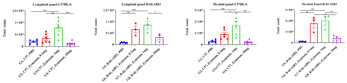
Increased CD45+ cells in peritoneal lavage observed in Zymosan induced acute peritonitis in C57BL/6 and B-hLAIR1 mice. Peritoneal lavage were collected 4 hours post zymosan injection for flow cytometry analysis for G1-G8. The total count of CD45+ cells were illustrated. Significantly increased CD45+ cells can be observed in both C57BL/6 mice and B-hLAIR1 mice administered 1 mg zymosan. For C57BL/6 mice, 0.1 mg zymosan also significantly increased CD45+ cells, though this is not observed in the B-hLAIR1 mice. One-way ANOVA followed by multiple comparison, all groups compared with each other. Values are expressed as mean ± SEM.
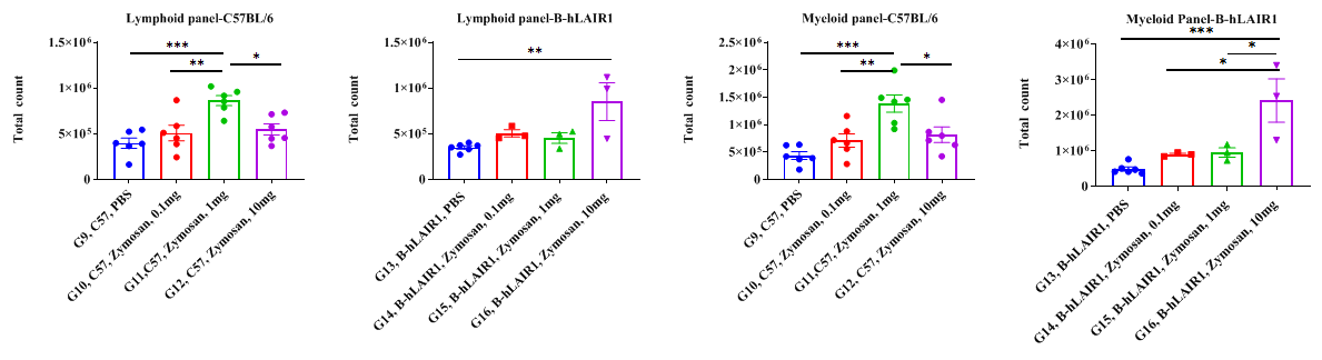
Increased CD45+ cells in peritoneal lavage observed in Zymosan induced sub-acute peritonitis in C57BL/6 and B-hLAIR1 mice. Peritoneal lavage were collected 3 days post zymosan injection for flow cytometry analysis for G9-G16. The total count of CD45+ cells were illustrated. Significantly increased CD45+ cells can be observed in both C57BL/6 mice and B-hLAIR1 mice administered 10 mg zymosan-D. For B-hLAIR1 mice, 1 mg zymosan, 10 mg zymosan also significantly increased CD45+ cells, though this is not observed in the C57BL/6 mice. One-way ANOVA followed by multiple comparison, all groups compared with each other. Values are expressed as mean ± SEM.
