

C57BL/6-Pdcd1tm1(PDCD1)Bcgen Cd274tm1(CD274)Bcgen/Bcgen • 120522
| Product name | B-hPD-1/hPD-L1 mice |
|---|---|
| Catalog number | 120522 |
| Strain name | C57BL/6-Pdcd1tm1(PDCD1)Bcgen Cd274tm1(CD274)Bcgen/Bcgen |
| Strain background | C57BL/6 |
| NCBI gene ID | 18566,16423 (Human) |
| Aliases | PDCD1 (Programmed death-1,as known as PD-1) ;CD274 (Programmed cell death ligand-1,also known as B7-H1, PD-L1) |
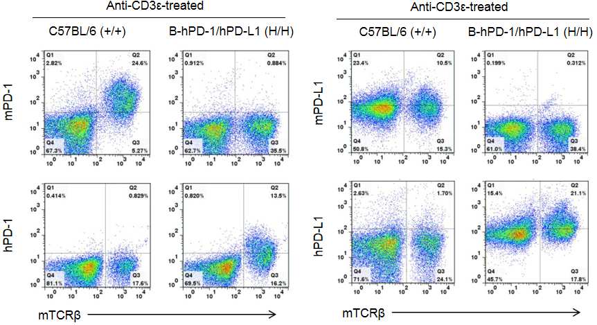
Strain specific PD1 and PD-L1 expression analysis in homozygous B-hPD-1/hPD-L1 mice by flow cytometry. Splenocytes were collected from WT and homozygous B-hPD-1/hPD-L1 (H/H) mice stimulated with anti-CD3ε in vivo , and analyzed by flow cytometry with species-specific anti-PD-1 and anti-PD-L1 antibodies. Mouse PD-1 and PD-L1 were exclusively detectable in WT mice. Human PD-1 and PD-L1 were exclusively detectable in homozygous B-hPD-1/hPD-L1 mice but not in WT mice.
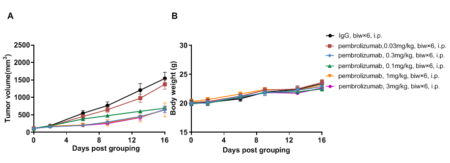
Antitumor activity of anti-human PD-1 antibody in B-hPD-1/hPD-L1 mice. (A) Anti-human PD-L1 antibody Pembrolizumab (in house) inhibited B-hPD-L1 MC38 plus tumor growth in B-hPD-1/hPD-L1 mice. Murine colon cancer B-hPD-L1 MC38 plus cells (5×10⁵) were subcutaneously implanted into homozygous B-hPD-1/hPD-L1 mice (male, 8-week-old, n=5). Mice were grouped when tumor volume reached approximately 80mm3, at which time they were treated with anti-human PD-1 antibody with doses and schedules indicated in panel. (B) Body weight changes during treatment. As shown in panel A, different doses of human PD-1 antibody inhibited tumor growth in different levels, demonstrating that the B-hPD-1/hPD-L1 mice provide a powerful preclinical model for in vivo evaluation of anti-human PD-1 antibodies. Values are expressed as mean ± SEM.
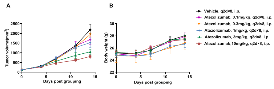
Antitumor activity of anti-human PD-L1 antibody in B-hPD-1/hPD-L1 mice. (A) Anti-human PD-L1 antibody atezolizumab (in house) inhibited B-hPD-L1 MC38 plus tumor growth in B-hPD-1/hPD-L1 mice. Murine colon cancer B-hPD-L1 MC38 plus cells (5×10⁵) were subcutaneously implanted into homozygous B-hPD-1/hPD-L1 mice (male, 5-week-old, n=5). Mice were grouped when tumor volume reached approximately 100 mm3, at which time they were treated with anti-human PD-L1 antibody with doses and schedules indicated in panel. (B) Body weight changes during treatment. As shown in panel A, different doses of human PD-L1 antibody inhibited tumor growth in different levels, demonstrating that the B-hPD-1/hPD-L1 mice provide a powerful preclinical model for in vivo evaluation of anti-human PD-L1 antibodies. Values are expressed as mean ± SEM.
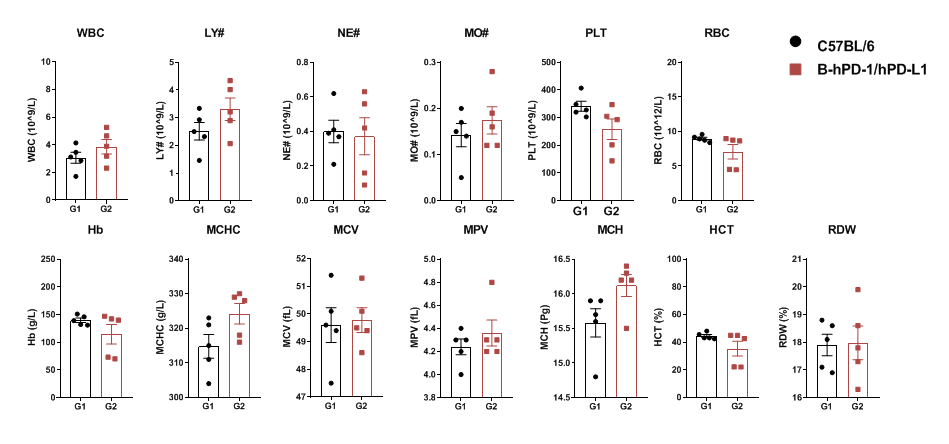
Complete blood count (CBC). Blood from female C57BL/6 and B-hPD-1/hPD-L1 mice (n = 5, 6 weeks-old) was collected and analyzed by CBC. There was no differences among any measurement between C57BL/6 and B-hPD-1/hPD-L1 mice.
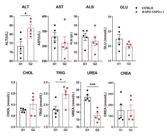
Blood chemistry tests of B-hPD-1/hPD-L1 mice. Serum from the C57BL/6 and B-hPD-1/hPD-L1 mice (n=3, 6 week-old) was collected and analyzed for levels of biochemistry. There was no differences on AST, ALB, GLU, CHOL, CREA measurement between C57BL/6 and B-hPD-1/hPD-L1,. ALT, TRIG, UREA have significant changes compared to wild-type mice. Values are expressed as mean ± SEM.
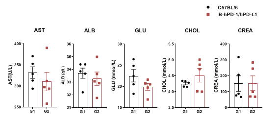
Blood chemistry tests of B-hPD-1/hPD-L1 mice. Serum from the C57BL/6 and B-hPD-1/hPD-L1 mice (n = 3, 6 week-old) was collected and analyzed for levels of biochemistry. There was no differences on AST, ALB, GLU, CHOL, CREA measurement between C57BL/6 and B-hPD-1/hPD-L1. Values are expressed as mean ± SEM.
