

BALB/c-Tnfrsf1btm1(TNFRSF1B)Bcgen/Bcgen • 112644
| Product name | B-hTNFR2 mice(C) |
|---|---|
| Catalog number | 112644 |
| Strain name | BALB/c-Tnfrsf1btm1(TNFRSF1B)Bcgen/Bcgen |
| Strain background | BALB/cCrSlcNifdc |
| NCBI gene ID | 21938 |
| Aliases | p75; TBPII; TNFBR; TNFR2; CD120b; TNFR1B; TNFR80; TNF-R75; p75TNFR; TNF-R-II |
| Application | For example, This product is used for pharmacodynamics and safety evaluation of cancer. |
on this page
Gene targeting strategy for B-hTFNR2 mice(C). The exons 2-6 of mouse Tfnr2 gene that encodes the extracellular domain was replaced by human TFNR2 exons 2-6 in B-hTFNR2 mice(C).
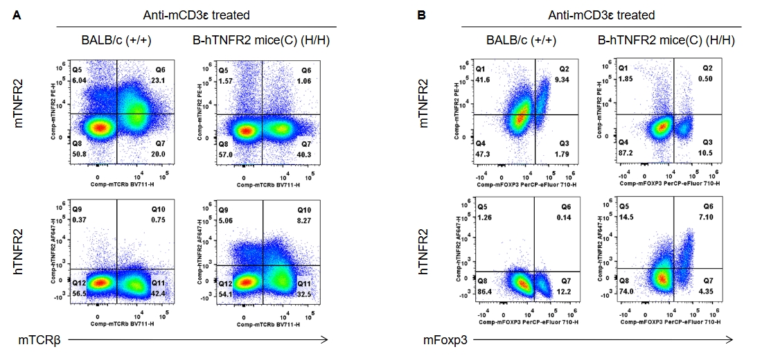
Strain specific TNFR2 expression analysis in wild-type and B-hTNFR2 mice(C) by flow cytometry. Human TNFR2 was exclusively detectable on T and Treg cells of homozygous B-hTNFR2 mice (C).
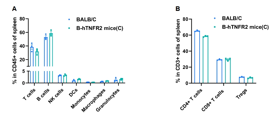
Frequency of leukocyte subpopulations in spleen by flow cytometry. Splenocytes were isolated from wild-type C57BL/6 mice and homozygous B-hTNFR2 mice(C) (n=3, 7-week-old, female). A. Flow cytometry analysis of the splenocytes was performed to assess the frequency of leukocyte subpopulations. B. Frequency of T cell subpopulations. Percentages of T cells, B cells, NK cells, dendritic cells, granulocytes, monocytes, macrophages, CD4+ T cells, CD8+ T cells and Tregs in B-hTNFR2 mice(C) were similar to those in C57BL/6 mice, demonstrating that humanization of TNFR2 does not change the frequency or distribution of these cell types in spleen. The frequency of leukocyte subpopulations in lymph node and blood of B-hTNFR2 mice(C) were also comparable to wild-type C57BL/6 mice (Data not shown). Values are expressed as mean ± SEM. Significance was determined by two-way ANOVA test. *P < 0.05, **P < 0.01, ***p < 0.001.
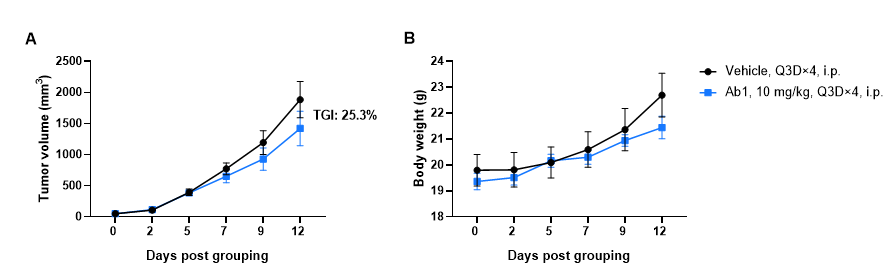
Antitumor activity of anti-human TNFR2 antibody in B-hTNFR2 mice(C). (A) Anti-human TNFR2 antibody inhibited CT26.WT tumor growth in B-hTNFR2 mice(C). Murine colon cancer CT26.WT cells were subcutaneously implanted into homozygous B-hTNFR2 mice(C) (female, 7-week-old, n=6). Mice were grouped when tumor volume reached approximately 40-60 mm3, at which time they were intraperitoneally injected with anti-human TNFR2 antibody indicated in panel. (B) Body weight changes during treatment. As shown in panel A, anti-human TNFR2 antibody was efficacious in controlling tumor growth in B-hTNFR2 mice(C), demonstrating that the B-hTNFR2 mice(C) provide a powerful preclinical model for in vivo evaluation of anti-human TNFR2 antibodies. Values are expressed as mean ± SEM. (The hTNFR2 Ab1 was provided by the client)
