

C57BL/6-Vegfatm1(VEGFA)Bcgen/Bcgen • 110822
| Product name | B-hVEGFA mice |
|---|---|
| Catalog number | 110822 |
| Strain name | C57BL/6-Vegfatm1(VEGFA)Bcgen/Bcgen |
| Strain background | C57BL/6 |
| Aliases | VEGFA (MVCD1, VEGF, VPF) |
Gene targeting strategy for B-hVEGFA mice. The exons 1-8 of mouse Vegfa gene that encode the full-length protein were replaced by human VEGFA exons 1-8 in B-hVEGFA mice.
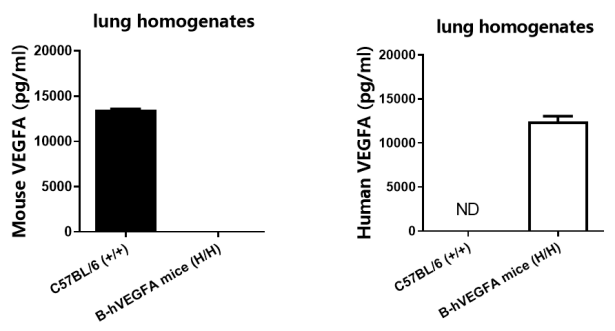
Strain specific VEGFA expression analysis in homozygous B-hVEGFA mice by ELISA. Mouse lung homogenates was collected from wild-type mice (+/+) and B-hVEGFA mice (H/H), and analyzed by ELISA with species-specific VEGFA kit. Mouse VEGFA was detectable in wild-type mice. Human VEGFA was exclusively detectable in B-hVEGFA mice but not in wild-type mice.
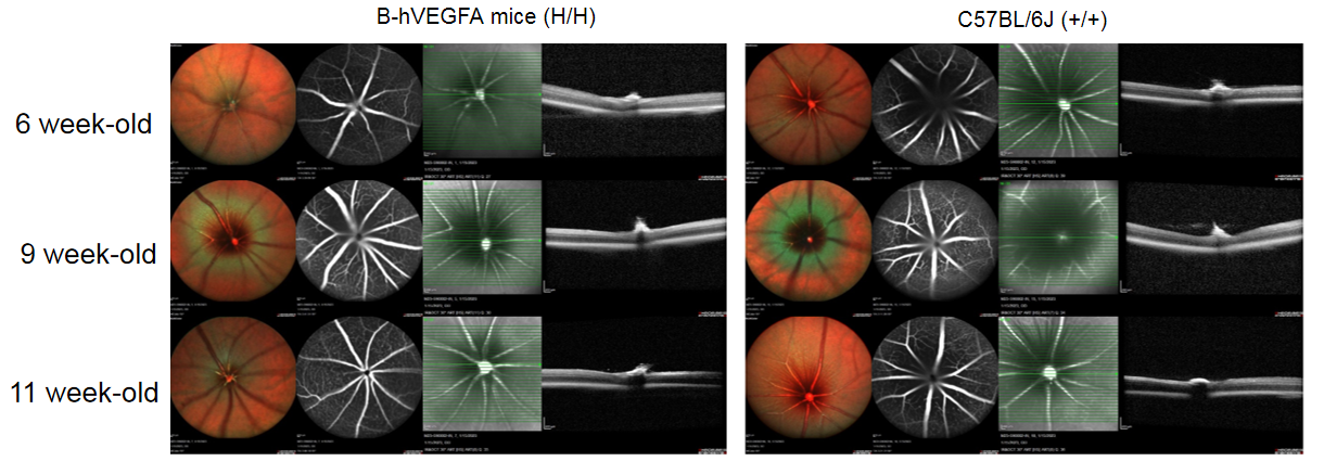
Fundus photographs of B-hVEGFA mice. Fundus images shows that the fundus morphology of B-hVEGFA mice has no significant change compared with wild-type mice(male, n=3).
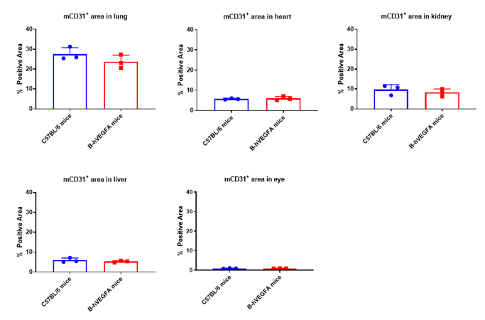
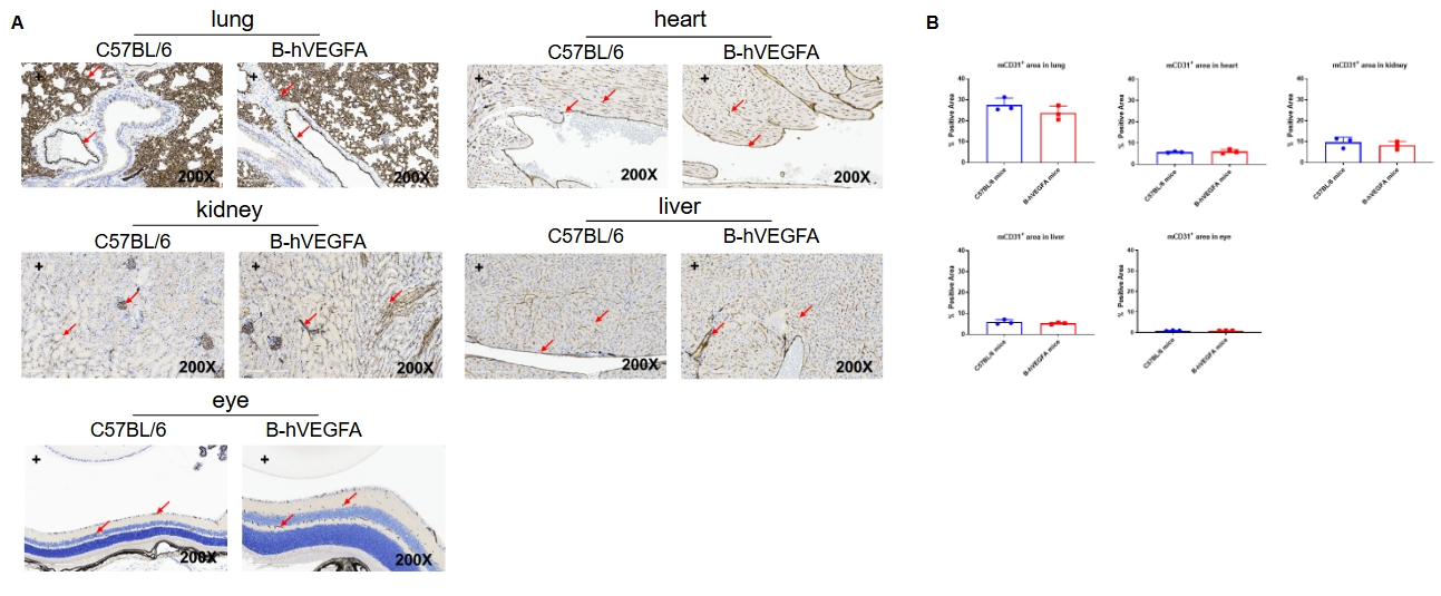
We investigated by immunohistochemistry the expression of mCD31 in the lung, heart, kidney, liver, and eye of C57BL/6 mice and B-hVEGFA mice (female, 8 week-old, n=3). The results showed that there was no significant difference in the proportion of mCD31+ between C57BL/6 mice and B-hVEGFA mice in lung, heart, kidney liver, and eye. It is suggested that VEGFA humanized has no effect on angiogenesis.
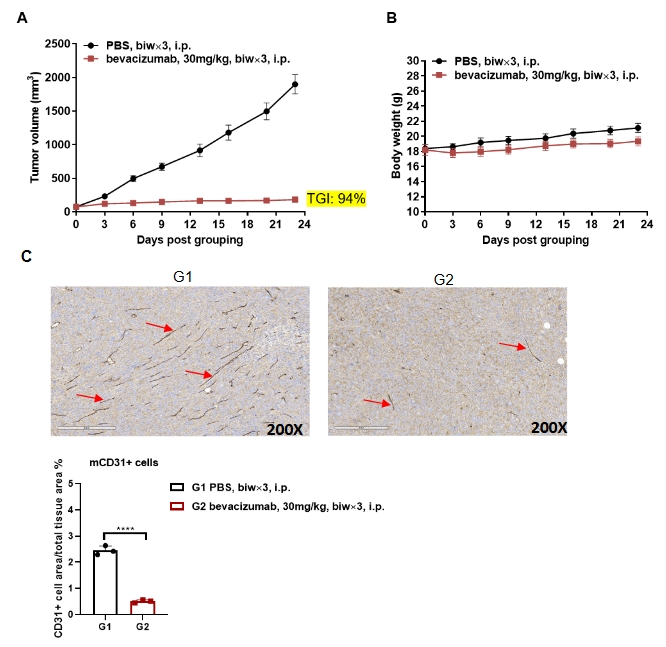
Antitumor activity of anti-human VEGFA antibody in B-hVEGFA mice. (A) Anti-human VEGFA antibody inhibited B-hVEGFA MC38 tumor growth in B-hVEGFA mice. Murine colon cancer B-hVEGFA MC38 cells were subcutaneously implanted into homozygous B-hVEGFA mice (female, 6-7-week-old, n=6). Mice were grouped when tumor volume reached approximately 100 mm3, at which time they were treated with anti-human VEGFA antibody bevacizumab and schedules indicated in panel A. (B) Body weight changes during treatment. (C) Proportion of CD31+ cell area of total area analyzed in tumor tissue. As shown in panel A, anti-human VEGFA antibody were efficacious in controlling tumor growth in B-hVEGFA mice. As shown in panel C, anti-human VEGFA antibody bevacizumab inhibits vascular endothelial cell proliferation, demonstrating that the B-hVEGFA mice provide a powerful preclinical model for in vivo evaluation of anti-human VEGFA antibody. Values are expressed as mean ± SEM.
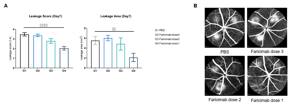
In vivo efficacy of anti-VEGFA and ANG2 bispecific antibody in B-hVEGFA mice. B-hVEGFA mice which established a choroidal neovascularizations (CNVs) model by laser treatment were randomly divided into four groups (n=4/group, female, 6-7 weeks). PBS (as a control) and bispecific antibody Faricimab targeted both VEGFA and ANG2 were administered to the mice individually (ivt, various dosages). All animals underwent FFA (fundus fluorescein angiography) on Day 7 to quantify the FFA scores (ranging from Grade I to IV,) and lesion area. (A) The leakage score and leakage area of B-hVEGFA mice after Faricimab treatment. (B) The FFA images of B-hVEGFA mice after Faricimab treatment on Day 7. Values are expressed as mean ± SEM, *P < 0.05, **P < 0.01: Faricimab (dose 1, 2, 3) vs PBS (One-Way ANOVA). Note: All the co-validation data were provided by pharmalegacy.
