C57BL/6-Icostm7(ICOS)Bcgen/Bcgen • 112105
| Product name | B-hICOS mice |
|---|---|
| Catalog number | 112105 |
| Strain name | C57BL/6-Icostm7(ICOS)Bcgen/Bcgen |
| Strain background | C57BL/6 |
| Aliases | ICOS, AILIM, CD278, CVID1, inducible T cell costimulator |
Gene targeting strategy for B-hICOS mice. The exons 1-5 of mouse Icos gene that encode the full-length protein were replaced by human ICOS exons 1-5 in B-hICOS mice.
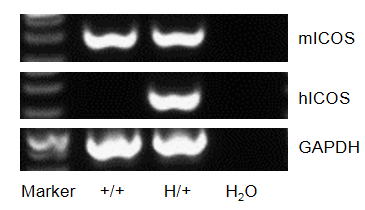
Strain specific analysis of ICOS gene expression in wild type (WT) mice and B-hICOS mice by RT-PCR. Mouse Icos mRNA was detectable in thymus of WT mice (+/+) and heterozygous B-hICOS mice (H/+). Human ICOS mRNA was detectable only in heterozygous B-hICOS mice (H/+) but not in WT mice (+/+). The positive band was confirmed to be correct by sequencing.
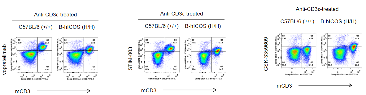
Strain specific ICOS expression analysis in homozygous B-hICOS mice by flow cytometry. Splenocytes were collected from wild-type (WT) mice (+/+) and homozygous B-hICOS mice (H/H) stimulated with anti-CD3ε in vivo and analyzed by flow cytometry with vopratelimab (in house), STIM-003 (in house) and GSK-3359609 (in house). ICOS was detectable in WT mice (+/+) and homozygous B-hICOS mice (H/H) due to the cross-reactivity of antibodies. Human ICOS was exclusively detectable in homozygous B-hICOS mice (H/H) but not in WT mice (+/+).
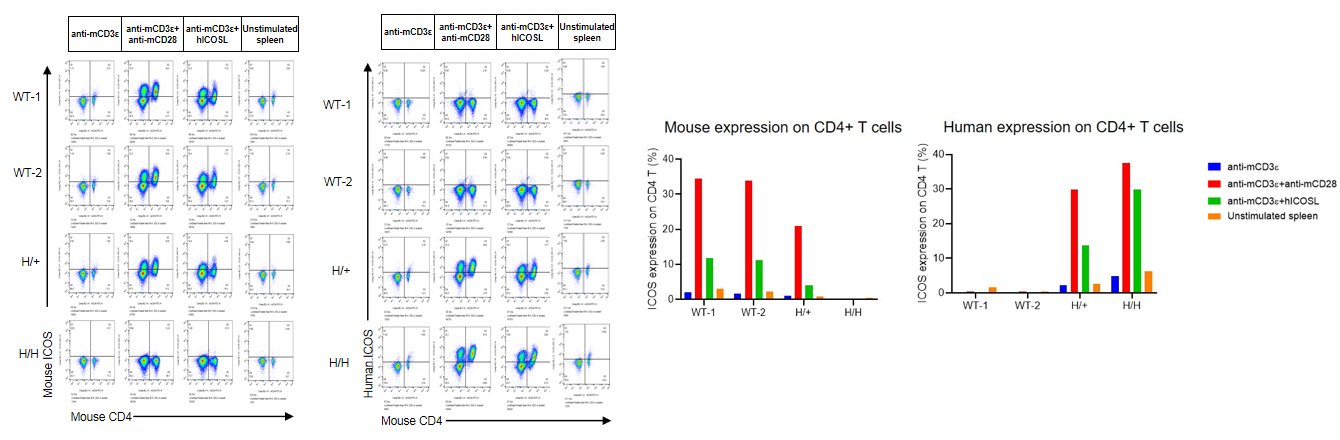
Mouse spleen CD4/CD8 ICOS expression and proliferation by flow cytometry. Splenocytes from wild-type (WT) mice (+/+), heterozygous B-hICOS mice (H/+) and homozygous B-hICOS mice (H/H) were stained by CellTrace Violet staining solution, and incubated for 72h in the presence of anti-mCD3ε, or anti-mCD3ε and anti-mCD28, or anti-mCD3ε and human ICOSL recombinant protein. The expression and proliferation of CD4+ T cells and CD8+ T cells were measured by flow cytometry. Mouse ICOS was detectable in WT mice (+/+) and heterozygous B-hICOS mice (H/+). Human ICOS was exclusively detectable in heterozygous B-hICOS mice (H/+) and homozygous B-hICOS mice (H/H) but not in WT mice (+/+). The data was obtained from a partner.
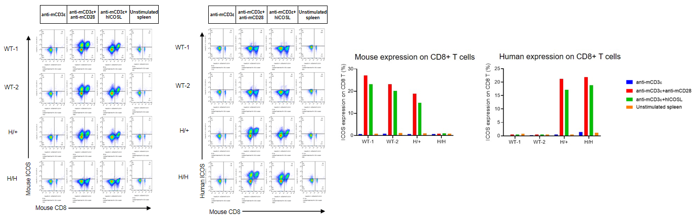
Mouse spleen CD4/CD8 ICOS expression and proliferation by flow cytometry. Splenocytes from wild-type (WT) mice (+/+), heterozygous B-hICOS mice (H/+) and homozygous B-hICOS mice (H/H) were stained by CellTrace Violet staining solution, and incubated for 72h in the presence of anti-mCD3ε, or anti-mCD3ε and anti-mCD28, or anti-mCD3ε and human ICOSL recombinant protein. The expression and proliferation of CD4+ T cells and CD8+ T cells were measured by flow cytometry. Mouse ICOS was detectable in WT mice (+/+) and heterozygous B-hICOS mice (H/+). Human ICOS was exclusively detectable in heterozygous B-hICOS mice (H/+) and homozygous B-hICOS mice (H/H) but not in WT mice (+/+). The data was obtained from a partner.
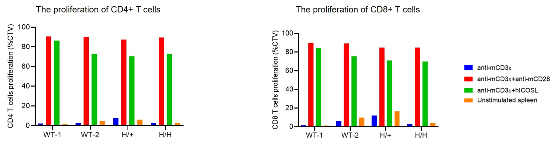
Mouse spleen CD4/CD8 ICOS expression and proliferation by flow cytometry. Splenocytes from wild-type (WT) mice (+/+), heterozygous B-hICOS mice (H/+) and homozygous B-hICOS mice (H/H) were stained by CellTrace Violet staining solution, and incubated for 72h in the presence of anti-mCD3ε, or anti-mCD3ε and anti-mCD28, or anti-mCD3ε and human ICOSL recombinant protein. The expression and proliferation of CD4+ T cells and CD8+ T cells were measured by flow cytometry. The CD4+ T cells and CD8+ T cells activation in heterozygous B-hICOS mice (H/+) and homozygous B-hICOS mice (H/H) was upregulated by anti-mCD3ε and human ICOSL recombinant protein, similar to the activation by anti-mCD3ε and anti-mCD28. This demonstrates that introduction of human ICOS in place of its mouse counterpart does not affect T cell activation. The data was obtained from a partner.