

C57BL/6-Il2tm1(IL2)Bcgen Il2ratm1(IL2RA) Bcgen ll2rbtm2(IL2RB)Bcgen ll2rgtm2(IL2RG)Bcgen/Bcgen • 112748
| Product name | B-hIL2/hIL2RA/hIL2RB/hIL2RG mice |
|---|---|
| Catalog number | 112748 |
| Strain name | C57BL/6-Il2tm1(IL2)Bcgen Il2ratm1(IL2RA) Bcgen ll2rbtm2(IL2RB)Bcgen ll2rgtm2(IL2RG)Bcgen/Bcgen |
| Strain background | C57BL/6 |
| Aliases | IL2 also known as TCGF, lymphokineIL2RA also known as CD25, IDDM10, IL2R, IMD41, TCGFR, p55IL2RB also known as CD122.IL2RG also known as CD132, SCIDX, IL-2RG |
Gene targeting strategy for B-hIL2/hIL2RA/hIL2RB/hIL2RG mice. The mouse Il2 gene that encodes the full coding sequence was replaced by human IL2 full coding sequence. The mouse Il2ra gene that encodes the extracellular domain was replaced by human IL2RA counterpart gene sequences. The mouse Il2rb gene that encodes the extracellular domain was replaced by human IL2RB counterpart gene sequences. The mouse Il2rg gene that encodes the full coding region sequences was replaced by human IL2RG counterpart gene sequences.
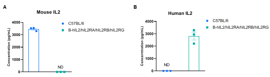
Strain specific IL2 expression analysis in homozygous B-hIL2/hIL2RA/IL2RB/IL2RG mice by ELISA. Serum were collected from wild-type mice C57BL/6 mice and homozygous B-hIL2/hIL2RA/IL2RB/IL2RG mice stimulated with anti-mCD3e antibody (7.5 μg/mice, i.p.) and anti-CD28 antibody (5 μg/mice, i.p.) in vivo for 2 h, and analyzed by ELISA with species-specific IL2 ELISA kit. (A) Mouse IL2 was only detectable in wild-type mice. (B) Human IL2 was exclusively detectable in homozygous mice but not in wild-type mice. Mice used in the experiment: male, 6-week-old, n=3/group.
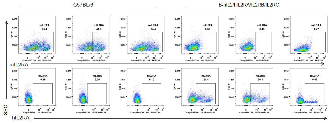
Strain specific IL2RA expression analysis in homozygous B-hIL2/hIL2RA/IL2RB/IL2RG mice by flow cytometry. Splenocytes were collected from wild-type C57BL/6 mice and homozygous B-hIL2/hIL2RA/IL2RB/IL2RG mice stimulated with anti-CD3e antibody in vivo (7.5 μg/mice, i.p.), and analyzed by flow cytometry with species-specific anti-IL2RA antibody. mIL2RA was only detectable in wild-type mice, and hIL2RA was exclusively detectable in T cells in homozygous B-hIL2/hIL2RA/IL2RB/IL2RG mice. Mice used in the experiment: male, 8-week-old, n=3/group.
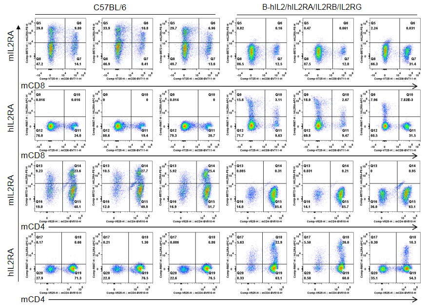
Strain specific IL2RA expression analysis in homozygous B-hIL2/hIL2RA/IL2RB/IL2RG by flow cytometry. Splenocytes were collected from wild-type C57BL/6 mice and homozygous B-hIL2/hIL2RA/IL2RB/IL2RG mice stimulated with anti-CD3e antibody in vivo (7.5 μg/mice, i.p.), and analyzed by flow cytometry with species-specific anti-IL2RA antibody. mIL2RA was only detectable in wild-type mice, and hIL2RA was exclusively detectable in CD4+ T and CD8+ T cells in homozygous B-hIL2/hIL2RA/IL2RB/IL2RG mice. Mice used in the experiment: male, 8-week-old, n=3/group.
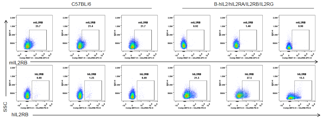
Strain specific IL2RB expression analysis in homozygous B-hIL2/hIL2RA/IL2RB/IL2RG mice by flow cytometry. Splenocytes were collected from wild-type C57BL/6 mice and homozygous B-hIL2/hIL2RA/IL2RB/IL2RG mice stimulated with anti-CD3e antibody in vivo (7.5 μg/mice, i.p.), and analyzed by flow cytometry with species-specific anti-IL2RB antibody. mIL2RB was only detectable in wild-type mice, and hIL2RB was exclusively detectable in T cells in homozygous B-hIL2/hIL2RA/IL2RB/IL2RG mice. Mice used in the experiment: male, 8-week-old, n=3/group.
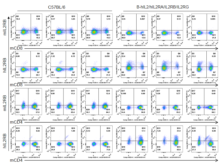
Strain specific IL2RB expression analysis in homozygous B-hIL2/hIL2RA/IL2RB/IL2RG by flow cytometry. Splenocytes were collected from wild-type C57BL/6 mice and homozygous B-hIL2/hIL2RA/IL2RB/IL2RG mice stimulated with anti-CD3e antibody in vivo (7.5 μg/mice, i.p.), and analyzed by flow cytometry with species-specific anti-IL2RB antibody. mIL2RB was only detectable in wild-type mice, and hIL2RB was exclusively detectable in CD4+ T and CD8+ T cells in homozygous B-hIL2/hIL2RA/IL2RB/IL2RG mice. Mice used in the experiment: male, 8-week-old, n=3/group.
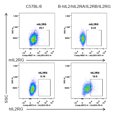
Strain specific IL2RG expression analysis in homozygous B-hIL2/hIL2RA/IL2RB/IL2RG by flow cytometry. Splenocytes were collected from wild-type C57BL/6 mice and homozygous B-hIL2/hIL2RA/IL2RB/IL2RG mice, and cultured in 96-well plates pre-coated with 2 μg/mL anti-mCD3e and 5 μg/mL anti-mCD28 antibody for 24 h, and then cells were harvested and analyzed by flow cytometry with species-specific anti-IL2RG antibody. mIL2RG was only detectable in wild-type mice, while hIL2RG was exclusively detectable in T cells in homozygous B-hIL2/hIL2RA/IL2RB/IL2RG mice. Mice used in the experiment: male, 8-week-old, 1 mice/group.
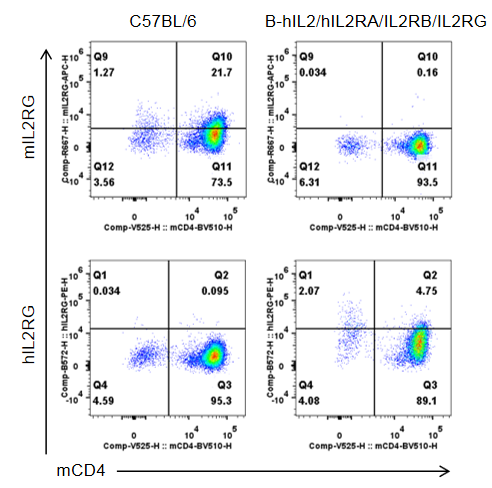
Strain specific IL2RG expression analysis in homozygous B-hIL2/hIL2RA/IL2RB/IL2RG by flow cytometry. Splenocytes were collected from wild-type C57BL/6 mice and homozygous B-hIL2/hIL2RA/IL2RB/IL2RG mice, and cultured in 96-well plates pre-coated with 2 μg/mL anti-mCD3e and 5 μg/mL anti-mCD28 antibody for 24 h, and then cells were harvested and analyzed by flow cytometry with species-specific anti-IL2RG antibody. mIL2RG was only detectable in wild-type mice, while hIL2RG was exclusively detectable in CD4+ T cells in homozygous B-hIL2/hIL2RA/IL2RB/IL2RG mice. Mice used in the experiment: male, 8-week-old, 1 mice/group.
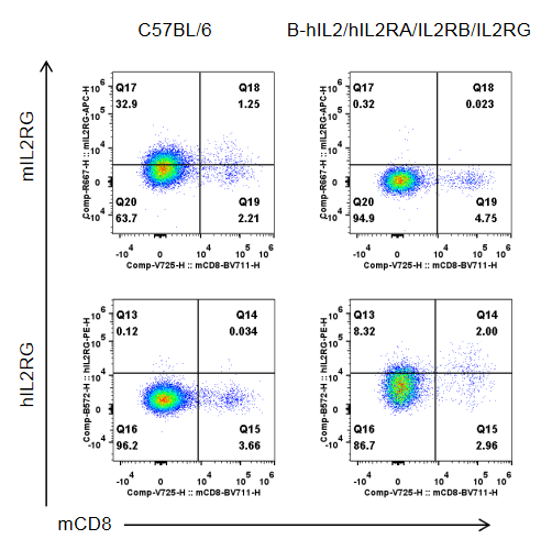
Strain specific IL2RG expression analysis in homozygous B-hIL2/hIL2RA/IL2RB/IL2RG by flow cytometry. Splenocytes were collected from wild-type C57BL/6 mice and homozygous B-hIL2/hIL2RA/IL2RB/IL2RG mice, and cultured in 96-well plates pre-coated with 2 μg/mL anti-mCD3e and 5 μg/mL anti-mCD28 antibody for 24 h, and then cells were harvested and analyzed by flow cytometry with species-specific anti-IL2RG antibody. mIL2RG was only detectable in wild-type mice, while hIL2RG was exclusively detectable in CD8+ T cells in homozygous B-hIL2/hIL2RA/IL2RB/IL2RG mice. Mice used in the experiment: male, 8-week-old, 1 mice/group.
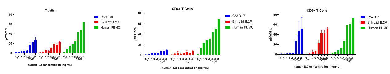
Intracellular phosphorylation of STAT5 analysis in splenocytes by flow cytometry. Splenocytes were harvested and analyzed for pSTAT5 induction in the T cell subtypes of wild type C57BL/6 mice and B-hIL2/hIL2RA/hIL2RB/hIL2RG mice. IL2 induced pSTAT5 in T cells with a dose dependent manner. At 10 ng/mL concentration, human IL2 induced higher pSTAT5 in T cells of B-hIL2/hIL2RA/hIL2RB/hIL2RG mice than that in wild type C57BL/6 mice.
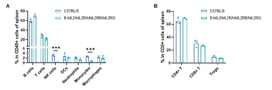
Frequency of leukocyte subpopulations in spleen by flow cytometry. Splenocytes were isolated from male wild-type C57BL/6 mice (n=3, 8-week-old) and homozygous B-hIL2/hIL2RA/IL2RB/IL2RG mice (n=3, 9-week-old). A. Flow cytometry analysis of the splenocytes was performed to assess the frequency of leukocyte subpopulations. B. Frequency of T cell subpopulations. Percentages of T cells, B cells, dendritic cells, neutrophils, macrophages, CD4+ T cells, CD8+ T cells and Tregs in B-hIL2/hIL2RA/IL2RB/IL2RG mice were similar to those in C57BL/6 mice. NK cells and monocytes decreased in humanized mice. Values are expressed as mean ± SD. Significance was determined by two-way ANOVA test. *P < 0.05, **P < 0.01, ***p < 0.001.
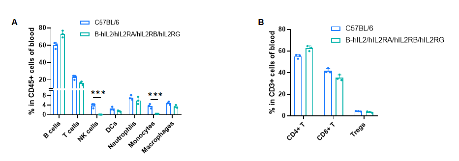
Frequency of leukocyte subpopulations in blood by flow cytometry. Blood cells were isolated from male wild-type C57BL/6 mice (n=3, 8-week-old) and homozygous B-hIL2/hIL2RA/IL2RB/IL2RG mice (n=3, 9-week-old). A. Flow cytometry analysis of the blood cells was performed to assess the frequency of leukocyte subpopulations. B. Frequency of T cell subpopulations. Percentages of T cells, B cells, dendritic cells, neutrophils, macrophages, CD4+ T cells, CD8+ T cells and Tregs in B-hIL2/hIL2RA/IL2RB/IL2RG mice were similar to those in C57BL/6 mice. NK cells and monocytes decreased in humanized mice. Values are expressed as mean ± SD. Significance was determined by two-way ANOVA test. *P < 0.05, **P < 0.01, ***p < 0.001.
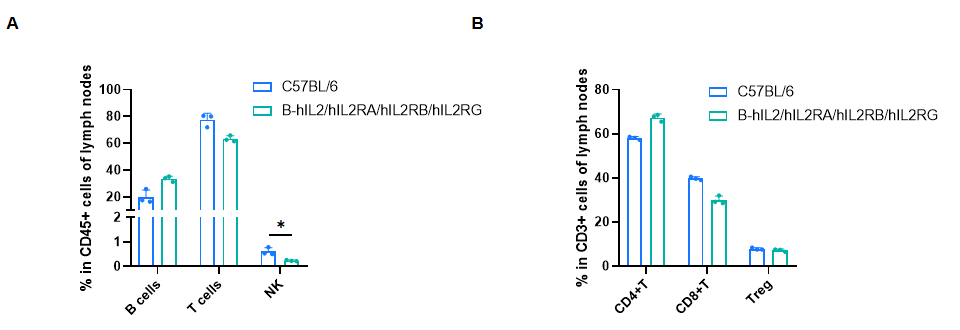
Frequency of leukocyte subpopulations in lymph nodes by flow cytometry. Lymph nodes cells were isolated from male wild-type C57BL/6 mice (n=3, 8-week-old) and homozygous B-hIL2/hIL2RA/IL2RB/IL2RG mice (n=3, 9-week-old). A. Flow cytometry analysis of the lymph nodes cells was performed to assess the frequency of leukocyte subpopulations. B. Frequency of T cell subpopulations. Percentages of T cells, B cells, CD4+ T cells, CD8+ T cells and Tregs in B-hIL2/hIL2RA/IL2RB/IL2RG mice were similar to those in C57BL/6 mice. NK cells decreased in humanized mice. Values are expressed as mean ± SD. Significance was determined by two-way ANOVA test. *P < 0.05, **P < 0.01, ***p < 0.001.
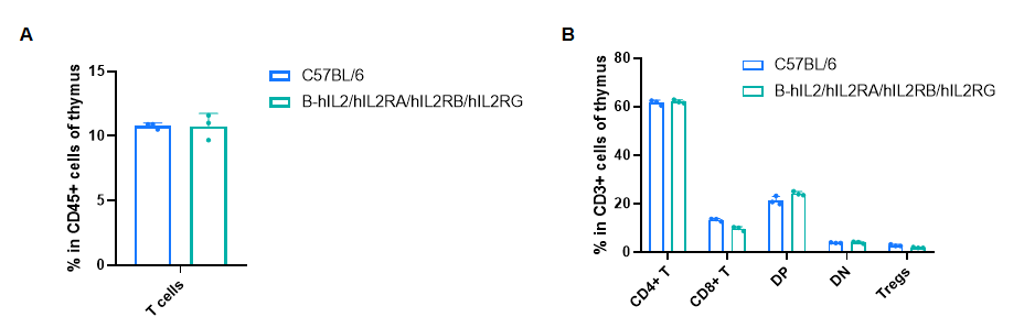
Frequency of leukocyte subpopulations in thymus by flow cytometry. Thymus cells were isolated from male wild-type C57BL/6 mice (n=3, 8-week-old) and homozygous B-hIL2/hIL2RA/IL2RB/IL2RG mice (n=3, 9-week-old). A. Flow cytometry analysis of the thymus cells was performed to assess the frequency of leukocyte subpopulations. B. Frequency of T cell subpopulations. Percentages of T cells, CD4+ T cells, CD8+ T cells and Tregs in B-hIL2/hIL2RA/IL2RB/IL2RG mice were similar to those in C57BL/6 mice. Values are expressed as mean ± SD. Significance was determined by two-way ANOVA test. *P < 0.05, **P < 0.01, ***p < 0.001
